3
views
0
recommends
+1 Recommend
1 collections
    0
    shares
      • Record: found
      • Abstract: found
      • Article: found
      Is Open Access

      DECTNet: Dual Encoder Network combined convolution and Transformer architecture for medical image segmentation

      research-article
      1 , 1 , 1 , * , , 2
      PLOS ONE
      Public Library of Science

      Read this article at

      Bookmark
          There is no author summary for this article yet. Authors can add summaries to their articles on ScienceOpen to make them more accessible to a non-specialist audience.

          Abstract

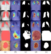
          Automatic and accurate segmentation of medical images plays an essential role in disease diagnosis and treatment planning. Convolution neural networks have achieved remarkable results in medical image segmentation in the past decade. Meanwhile, deep learning models based on Transformer architecture also succeeded tremendously in this domain. However, due to the ambiguity of the medical image boundary and the high complexity of physical organization structures, implementing effective structure extraction and accurate segmentation remains a problem requiring a solution. In this paper, we propose a novel Dual Encoder Network named DECTNet to alleviate this problem. Specifically, the DECTNet embraces four components, which are a convolution-based encoder, a Transformer-based encoder, a feature fusion decoder, and a deep supervision module. The convolutional structure encoder can extract fine spatial contextual details in images. Meanwhile, the Transformer structure encoder is designed using a hierarchical Swin Transformer architecture to model global contextual information. The novel feature fusion decoder integrates the multi-scale representation from two encoders and selects features that focus on segmentation tasks by channel attention mechanism. Further, a deep supervision module is used to accelerate the convergence of the proposed method. Extensive experiments demonstrate that, compared to the other seven models, the proposed method achieves state-of-the-art results on four segmentation tasks: skin lesion segmentation, polyp segmentation, Covid-19 lesion segmentation, and MRI cardiac segmentation.

          Related collections

          Most cited references26

          • Record: found
          • Abstract: found
          • Article: not found

          A survey on deep learning in medical image analysis

          Deep learning algorithms, in particular convolutional networks, have rapidly become a methodology of choice for analyzing medical images. This paper reviews the major deep learning concepts pertinent to medical image analysis and summarizes over 300 contributions to the field, most of which appeared in the last year. We survey the use of deep learning for image classification, object detection, segmentation, registration, and other tasks. Concise overviews are provided of studies per application area: neuro, retinal, pulmonary, digital pathology, breast, cardiac, abdominal, musculoskeletal. We end with a summary of the current state-of-the-art, a critical discussion of open challenges and directions for future research.
            Bookmark
            • Record: found
            • Abstract: found
            • Article: not found

            CE-Net: Context Encoder Network for 2D Medical Image Segmentation

            Medical image segmentation is an important step in medical image analysis. With the rapid development of a convolutional neural network in image processing, deep learning has been used for medical image segmentation, such as optic disc segmentation, blood vessel detection, lung segmentation, cell segmentation, and so on. Previously, U-net based approaches have been proposed. However, the consecutive pooling and strided convolutional operations led to the loss of some spatial information. In this paper, we propose a context encoder network (CE-Net) to capture more high-level information and preserve spatial information for 2D medical image segmentation. CE-Net mainly contains three major components: a feature encoder module, a context extractor, and a feature decoder module. We use the pretrained ResNet block as the fixed feature extractor. The context extractor module is formed by a newly proposed dense atrous convolution block and a residual multi-kernel pooling block. We applied the proposed CE-Net to different 2D medical image segmentation tasks. Comprehensive results show that the proposed method outperforms the original U-Net method and other state-of-the-art methods for optic disc segmentation, vessel detection, lung segmentation, cell contour segmentation, and retinal optical coherence tomography layer segmentation.
              Bookmark
              • Record: found
              • Abstract: found
              • Article: not found

              WM-DOVA maps for accurate polyp highlighting in colonoscopy: Validation vs. saliency maps from physicians.

              We introduce in this paper a novel polyp localization method for colonoscopy videos. Our method is based on a model of appearance for polyps which defines polyp boundaries in terms of valley information. We propose the integration of valley information in a robust way fostering complete, concave and continuous boundaries typically associated to polyps. This integration is done by using a window of radial sectors which accumulate valley information to create WM-DOVA (Window Median Depth of Valleys Accumulation) energy maps related with the likelihood of polyp presence. We perform a double validation of our maps, which include the introduction of two new databases, including the first, up to our knowledge, fully annotated database with clinical metadata associated. First we assess that the highest value corresponds with the location of the polyp in the image. Second, we show that WM-DOVA energy maps can be comparable with saliency maps obtained from physicians' fixations obtained via an eye-tracker. Finally, we prove that our method outperforms state-of-the-art computational saliency results. Our method shows good performance, particularly for small polyps which are reported to be the main sources of polyp miss-rate, which indicates the potential applicability of our method in clinical practice.
                Bookmark

                Author and article information

                Contributors
                Role: InvestigationRole: MethodologyRole: VisualizationRole: Writing – original draft
                Role: Writing – review & editing
                Role: Supervision
                Role: Validation
                Role: Editor
                Journal
                PLoS One
                PLoS One
                plos
                PLOS ONE
                Public Library of Science (San Francisco, CA USA )
                1932-6203
                2024
                4 April 2024
                : 19
                : 4
                : e0301019
                Affiliations
                [1 ] Department of Control Science and Engineering, Harbin Institute of Technology, Harbin, Heilongjiang, China
                [2 ] Sergeant Schools of Army Academy of Armored Forces, Changchun, Jilin, China
                Vellore Institute of Technology: VIT University, INDIA
                Author notes

                Competing Interests: The authors have declared that no competing interests exist.

                Author information
                https://orcid.org/0009-0001-0757-8919
                Article
                PONE-D-23-34762
                10.1371/journal.pone.0301019
                10994332
                38573957
                0da16bfc-9537-468c-b82f-247c9b0bb57c
                © 2024 Li et al

                This is an open access article distributed under the terms of the Creative Commons Attribution License, which permits unrestricted use, distribution, and reproduction in any medium, provided the original author and source are credited.

                History
                : 27 October 2023
                : 9 March 2024
                Page count
                Figures: 14, Tables: 6, Pages: 25
                Funding
                The author(s) received no specific funding for this work.
                Categories
                Research Article
                Research and Analysis Methods
                Mathematical and Statistical Techniques
                Mathematical Functions
                Convolution
                Medicine and Health Sciences
                Clinical Medicine
                Signs and Symptoms
                Lesions
                Biology and Life Sciences
                Neuroscience
                Cognitive Science
                Cognitive Psychology
                Attention
                Biology and Life Sciences
                Psychology
                Cognitive Psychology
                Attention
                Social Sciences
                Psychology
                Cognitive Psychology
                Attention
                Medicine and Health Sciences
                Medical Conditions
                Infectious Diseases
                Viral Diseases
                Covid 19
                Biology and Life Sciences
                Anatomy
                Cardiovascular Anatomy
                Heart
                Cardiac Ventricles
                Medicine and Health Sciences
                Anatomy
                Cardiovascular Anatomy
                Heart
                Cardiac Ventricles
                Computer and Information Sciences
                Computer Architecture
                Research and Analysis Methods
                Imaging Techniques
                Medicine and Health Sciences
                Diagnostic Medicine
                Clinical Laboratory Sciences
                Transfusion Medicine
                Medicine and Health Sciences
                Hematology
                Transfusion Medicine
                Custom metadata
                All of the datasets employed in the paper are available from https://www.kaggle.com/datasets/lbl1993/skin-covid-19-polyp-cardiac-segmentataion-datasets.
                COVID-19

                Uncategorized
                Uncategorized

                Comments

                Comment on this article