6
views
0
recommends
+1 Recommend
0 collections
    0
    shares
      • Record: found
      • Abstract: found
      • Article: found
      Is Open Access

      Long-term effects of phacoemulsification and intraocular lens implantation in a patient with pathologic myopia and extremely long axial length : A case report

      case-report

      Read this article at

      Bookmark
          There is no author summary for this article yet. Authors can add summaries to their articles on ScienceOpen to make them more accessible to a non-specialist audience.

          Abstract

          Rationale:

          To report a rare case of phacoemulsification cataract surgery and intraocular lens implantation that improved visual acuity and capsular stability in a patient with pathologic myopia and axial length >38 mm.

          Patient concerns:

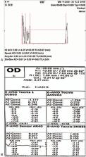
          A 51-year-old Chinese man with high myopia since childhood who had lost sight in his left eye at the age of 25 due to retinal detachment. He was referred for ophthalmological assessment due to decreased vision in the right eye, in which the best-corrected visual acuity at distance was hand motion.

          Diagnoses:

          The patient was diagnosed with cataract, high myopia, subluxated lens, and loose zonules in the right eye. The left eyeball showed atrophy.

          Interventions:

          The patient underwent uneventful phacoemulsification. An intraocular lens (Sensar AR40M) and capsular tension ring were implanted within the capsular bag. After surgery, the patient was given eye drops containing tobramycin and dexamethasone eye drops for 1 month and eye drops containing 0.1% sodium diclofenac for 2 months.

          Outcomes:

          There were no postoperative complications. During 1-year follow-up, uncorrected visual acuity was 20/80 and the manifest refraction was −2.50DS/−1.00DC∗80, with corrected distance visual acuity of 20/60. Cataract surgery maintained adequate vision for daily living.

          Lessons:

          Implantation of specific lens and capsular tension ring as well as prolonged use of non-steroidal anti-inflammatory drugs may help prevent capsular contraction and posterior capsule opacification in patients with pathologic myopia and extremely long axial length.

          Related collections

          Most cited references19

          • Record: found
          • Abstract: found
          • Article: not found

          Updates of pathologic myopia.

          Complications from pathologic myopia are a major cause of visual impairment and blindness, especially in east Asia. The eyes with pathologic myopia may develop loss of the best-corrected vision due to various pathologies in the macula, peripheral retina and the optic nerve. Despite its importance, the definition of pathologic myopia has been inconsistent. The refractive error or axial length alone often does not adequately reflect the 'pathologic myopia'. Posterior staphyloma, which is a hallmark lesion of pathologic myopia, can occur also in non-highly myopic eyes. Recently a revised classification system for myopic maculopathy has been proposed to standardize the definition among epidemiological studies. In this META-PM (meta analyses of pathologic myopia) study classification, pathologic myopia was defined as the eyes having chorioretinal atrophy equal to or more severe than diffuse atrophy. In addition, the advent of new imaging technologies such as optical coherence tomography (OCT) and three dimensional magnetic resonance imaging (3D MRI) has enabled the detailed observation of various pathologies specific to pathologic myopia. New therapeutic approaches including intravitreal injections of anti-vascular endothelial growth factor agents and the advance of vitreoretinal surgeries have greatly improved the prognosis of patients with pathologic myopia. The purpose of this review article is to provide an update on topics related to the field of pathologic myopia, and to outline the remaining issues which need to be solved in the future.
            Bookmark
            • Record: found
            • Abstract: found
            • Article: not found

            Histological changes of high axial myopia.

            J Jonas, L. Xu (2014)
            To describe pathological changes in the anatomy of highly myopic (axially elongated) eyes, enucleated globes were examined by light microscopy and ocular structures were measured histomorphometrically. These studies revealed that highly axially myopic eyes show continuous thinning of the sclera starting at or behind the equator with a maximal thinning at the posterior pole; a profound thinning of the choroid decreasing from ∼ 250 to <10 μm in extreme axial myopia, secondary macular defects in the Bruch's membrane associated with a complete loss of retinal pigment epithelium and choriocapillaris, and retinal photoreceptors; a Bruch's membrane of normal thickness in contrast to the profound thinning of the choroid and the sclera; an up to 10-fold elongation and thinning of the peripapillary scleral flange as anterior roof of the orbital cerebrospinal fluid space, and subsequently a retrobulbar extension of the cerebrospinal fluid space; an increased distance of the peripapillary arterial circle of Zinn-Haller to the optic disc border; an elongation and thinning of the lamina cribrosa with a subsequently decreased distance between the intraocular pressure compartment and the retrobulbar orbital cerebrospinal fluid pressure compartment; an increasing exposure of the peripheral posterior lamina cribrosa surface to the cerebrospinal fluid space, no longer buffered by the solid optic nerve tissue; and the development and enlargement of parapapillary gamma zone, in contrast to a myopia-independent parapapillary beta zone. These anatomical changes may be associated with high axial myopia-related complications such as an increased susceptibility of glaucomatous optic neuropathy and myopic retinopathy.
              Bookmark
              • Record: found
              • Abstract: found
              • Article: not found

              Proinflammatory status in the aqueous humor of high myopic cataract eyes.

              High myopia has long been recognized as an inflammation-related disease, and high myopic eyes are thought to have a proinflammatory internal microenvironment, which might predispose to the occurrence of certain inflammation-related complications such as fibrotic capsular contraction syndrome after cataract surgery. Therefore, the purpose of this study was to detect inflammatory cytokines expressed in the aqueous humor (AH) of high myopic cataract (HMC) patients. The cytokines were screened using a RayBio Human Cytokine Antibody Array in AH samples from 15 age-related cataract (ARC) patients and 15 HMC patients. Those detected by the screening assays were verified using a Bio-Plex Suspension Array System in AH samples from 35 ARC patients and 45 HMC patients. The cytokine antibody array showed that the expression level of interleukin-1 receptor antagonist (IL-1ra) in the AH was higher in ARC than in HMC, whereas opposite trends were found for monocyte chemoattractant protein-1 (MCP-1), regulated on activation, normal T-cell expressed and presumably secreted (RANTES), IL-8, platelet-derived growth factor-BB, and IL-6 (all P < 0.05). In the verification assay using the suspension cytokine array, only the expression levels of IL-1ra and MCP-1 were significantly different between the ARC and HMC groups (P = 0.014 and 0.038, respectively); these results were confirmed by western blot assays. Our results demonstrated that the expression of IL-1ra was significantly lower and the expression of MCP-1 was significantly higher in the AH of HMC than in ARC, suggestive of a proinflammatory status in the anterior chamber of HMC eyes.
                Bookmark

                Author and article information

                Journal
                Medicine (Baltimore)
                Medicine (Baltimore)
                MEDI
                Medicine
                Lippincott Williams & Wilkins (Hagerstown, MD )
                0025-7974
                1536-5964
                11 September 2020
                11 September 2020
                : 99
                : 37
                : e22081
                Affiliations
                Department of Ophthalmology, West China Hospital of Sichuan University, Chengdu, Sichuan Province, China.
                Author notes
                []Correspondence: Wei Fan, Department of Ophthalmology, West China Hospital of Sichuan University, Chengdu, Sichuan Province 610041, China (e-mail: fanwei55@ 123456yahoo.com ).
                Article
                MD-D-20-00598 22081
                10.1097/MD.0000000000022081
                7489675
                32925746
                06c81a3e-618a-4128-ab79-7a65c00e4fbb
                Copyright © 2020 the Author(s). Published by Wolters Kluwer Health, Inc.

                This is an open access article distributed under the Creative Commons Attribution License 4.0 (CCBY), which permits unrestricted use, distribution, and reproduction in any medium, provided the original work is properly cited. http://creativecommons.org/licenses/by/4.0

                History
                : 20 January 2020
                : 19 June 2020
                : 7 August 2020
                Categories
                5800
                Research Article
                Clinical Case Report
                Custom metadata
                TRUE

                capsular stability,capsular tension rings,cataract surgery,extremely long axial length,pathologic myopia

                Comments

                Comment on this article