0
views
0
recommends
+1 Recommend
0 collections
    0
    shares
      • Record: found
      • Abstract: found
      • Article: found
      Is Open Access

      Incidence and Risk Factors for Retinal Detachment Following Pediatric Cataract Surgery

      research-article

      Read this article at

      Bookmark
          There is no author summary for this article yet. Authors can add summaries to their articles on ScienceOpen to make them more accessible to a non-specialist audience.

          Abstract

          Background

          Retinal detachment is a major postsurgical threat in pediatric cataract surgery; however, the effect of axial length remains unclear. This study aimed to assess the relationship between axial length and detachment risk in vulnerable patients.

          Methods

          This retrospective cohort study analyzed 132 eyes of 84 pediatric cataract surgery patients aged <20 years old. Axial length was measured preoperatively, and the incidence of retinal detachment was recorded over a median follow-up of 4 years. Logistic regression analysis was used to examine the axial length-detachment relationship.

          Results

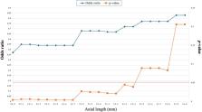
          Twenty eyes had postoperative retinal detachments. The median axial length was longer in the detachment group (23.6 mm) than in the non-detachment group (21.6 mm). Eyes with axial length ≤23.4 mm had 0.55-fold decreased odds of detachment compared to longer eyes. Preexisting myopia and glaucoma confer heightened risk. Approximately half of the patients retained some detachment risk eight years postoperatively.

          Conclusion

          Shorter eyes (axial length ≤23.4 mm) appear to be protected against pediatric retinal detachment after cataract surgery, whereas myopia, glaucoma, and axial elongation > 23.4 mm elevate the postoperative risk. Understanding these anatomical risk profiles requires surgical planning and follow-up care of children undergoing lensectomy.

          Plain language Summary

          This study investigated the protective role of a shorter axial length in preventing retinal detachment after pediatric cataract surgery. This highlights the correlation between smaller eye sizes and reduced detachment risk, emphasizing the need for careful consideration of anatomical factors in surgical planning and patient monitoring, particularly for patients with preexisting myopia or postoperative glaucoma.

          Graphical Abstract

          Related collections

          Most cited references41

          • Record: found
          • Abstract: found
          • Article: not found

          The impact of ocular blood flow in glaucoma.

          Two principal theories for the pathogenesis of glaucomatous optic neuropathy (GON) have been described--a mechanical and a vascular theory. Both have been defended by various research groups over the past 150 years. According to the mechanical theory, increased intraocular pressure (IOP) causes stretching of the laminar beams and damage to retinal ganglion cell axons. The vascular theory of glaucoma considers GON as a consequence of insufficient blood supply due to either increased IOP or other risk factors reducing ocular blood flow (OBF). A number of conditions such as congenital glaucoma, angle-closure glaucoma or secondary glaucomas clearly show that increased IOP is sufficient to lead to GON. However, a number of observations such as the existence of normal-tension glaucoma cannot be satisfactorily explained by a pressure theory alone. Indeed, the vast majority of published studies dealing with blood flow report a reduced ocular perfusion in glaucoma patients compared with normal subjects. The fact that the reduction of OBF often precedes the damage and blood flow can also be reduced in other parts of the body of glaucoma patients, indicate that the hemodynamic alterations may at least partially be primary. The major cause of this reduction is not atherosclerosis, but rather a vascular dysregulation, leading to both low perfusion pressure and insufficient autoregulation. This in turn may lead to unstable ocular perfusion and thereby to ischemia and reperfusion damage. This review discusses the potential role of OBF in glaucoma and how a disturbance of OBF could increase the optic nerve's sensitivity to IOP.
            Bookmark
            • Record: found
            • Abstract: found
            • Article: not found

            Anomalous posterior vitreous detachment: a unifying concept in vitreo-retinal disease.

            J. Sebag (2004)
            Posterior vitreous detachment (PVD) is the consequence of changes in the macromolecular structure of gel vitreous that result in liquefaction, concurrent with alterations in the extracellular matrix at the vitro-retinal interface that allow the posterior vitreous cortex to detach from the internal limiting lamina of the retina. Gel liquefaction that exceeds the degree of vitro-retinal dehiscence results in anomalous PVD (APVD). APVD varies in its clinical manifestations depending upon where in the fundus vitreo-retinal adhesion is strongest. At the periphery, APVD results in retinal tears and detachments. In the macula, APVD causes vitreo-macular traction syndrome, results in vitreoeschisis with macular pucker or macular holes, or contributes to some cases of diabetic macular edema. At the optic disc and retina, APVD causes vitreo-papillary traction and promotes retinal and optic disc neovascularization. Unifying the spectrum of vitreo-retinal diseases into the conceptual frame-work of APVD underscores that to more effectively treat, and ultimately prevent, these disorders it is necessary to replicate the two components of an innocuous PVD, i.e., gel liquefaction and vitreo-retinal dehiscence. Pharmacologic vitreolysis is designed to mitigate against APVD by chemically breaking down vitreous macromolecules and weakening vitro-retinal adhesion to safely detach the posterior vitreous cortex. This would not only facilitate surgery, but if performed early in the natural history of disease, it should prevent progressive disease.
              Bookmark
              • Record: found
              • Abstract: found
              • Article: not found

              Relationship of age, sex, and ethnicity with myopia progression and axial elongation in the correction of myopia evaluation trial.

              To identify the baseline factors independently related to 3-year myopia progression and axial elongation in COMET. A total of 469 children were enrolled, randomly assigned to progressive addition lenses with a + 2.00 diopter (D) addition or to single vision lenses and observed for 3 years. Eligible children were 6 to 11 years old, with spherical equivalent myopia of - 1.25 to - 4.50 D, bilaterally. The primary and secondary outcomes, myopia progression by cycloplegic autorefraction and axial elongation by A-scan ultrasonography, were measured annually. Multiple linear regression was used to adjust for covariates, including treatment. Younger baseline age (6-7 vs 11 years, 8 vs 11 years, and 9 vs 11 years, P<.001; 10 vs 11 years, P = .04), female sex (P = .01), and each ethnic group compared with African Americans (Asian, P = .02; Hispanic, P = .002; mixed, P = .002; white, P = .001) were independently associated with faster 3-year progression. Children aged 6 to 7 years had the fastest progression of all age groups, progressing by a mean (+/- SD) of 1.31 D +/- 0.13 more than children aged 11 years. Females progressed 0.16 D more than the males. Children of mixed, Hispanic, Asian, and white ethnicity progressed more than African American children by 0.49 D +/- 0.16, 0.33 D +/- 0.11, 0.32 D +/- 0.13, 0.27 D +/- 0.08, respectively. Age and ethnicity, but not sex, were independently associated with axial elongation. Among these myopic children, a 0.5 mm increase in axial length was associated with 1 D of myopia progression. Younger baseline age was the strongest factor independently associated with faster myopic progression and greater axial elongation at 3 years. African American children had less myopic progression and axial elongation than the other ethnic groups.
                Bookmark

                Author and article information

                Journal
                Clin Ophthalmol
                Clin Ophthalmol
                opth
                Clinical Ophthalmology (Auckland, N.Z.)
                Dove
                1177-5467
                1177-5483
                05 June 2024
                2024
                : 18
                : 1623-1636
                Affiliations
                [1 ]Pediatric Ophthalmology and Strabismus Division, King Khaled Eye Specialist Hospital , Riyadh, Saudi Arabia
                [2 ]Vitreoretinal Division, King Khaled Eye Specialist Hospital , Riyadh, Saudi Arabia
                [3 ]Faculty of Medicine, King Abdulaziz University , Jeddah, Saudi Arabia
                Author notes
                Correspondence: Gorka Sesma, Pediatric Ophthalmology and Strabismus Division, King Khaled Eye Specialist Hospital , Al Urubah Branche Road, West Building 2nd Floor, Riyadh, 11462, Saudi Arabia, Tel +966114849700, Fax +966114821908, Email gsesma@kkesh.med.sa
                [*]

                These authors contributed equally to this work

                Author information
                http://orcid.org/0000-0002-6125-3082
                http://orcid.org/0000-0002-4320-448X
                Article
                464005
                10.2147/OPTH.S464005
                11162630
                38855013
                bbb11788-f4a7-4bc8-b3a6-c3d193fd0723
                © 2024 Sabr et al.

                This work is published and licensed by Dove Medical Press Limited. The full terms of this license are available at https://www.dovepress.com/terms.php and incorporate the Creative Commons Attribution – Non Commercial (unported, v3.0) License ( http://creativecommons.org/licenses/by-nc/3.0/). By accessing the work you hereby accept the Terms. Non-commercial uses of the work are permitted without any further permission from Dove Medical Press Limited, provided the work is properly attributed. For permission for commercial use of this work, please see paragraphs 4.2 and 5 of our Terms ( https://www.dovepress.com/terms.php).

                History
                : 13 February 2024
                : 21 May 2024
                Page count
                Figures: 2, Tables: 8, References: 41, Pages: 14
                Funding
                Funded by: funding;
                There is no funding to report.
                Categories
                Original Research

                Ophthalmology & Optometry
                pediatric cataract surgery,axial length,retinal detachment,myopia,congenital glaucoma,congenital cataract

                Comments

                Comment on this article