36
views
0
recommends
+1 Recommend
0 collections
    0
    shares
      • Record: found
      • Abstract: found
      • Article: found
      Is Open Access

      Gene expression profiling reveals novel TGFβ targets in adult lung fibroblasts

      research-article

      Read this article at

      Bookmark
          There is no author summary for this article yet. Authors can add summaries to their articles on ScienceOpen to make them more accessible to a non-specialist audience.

          Abstract

          Background

          Transforming growth factor beta (TGFβ), a multifunctional cytokine, plays a crucial role in the accumulation of extracellular matrix components in lung fibrosis, where lung fibroblasts are considered to play a major role. Even though the effects of TGFβ on the gene expression of several proteins have been investigated in several lung fibroblast cell lines, the global pattern of response to this cytokine in adult lung fibroblasts is still unknown.

          Methods

          We used Affymetrix oligonucleotide microarrays U95v2, containing approximately 12,000 human genes, to study the transcriptional profile in response to a four hour treatment with TGFβ in control lung fibroblasts and in fibroblasts from patients with idiopathic and scleroderma-associated pulmonary fibrosis. A combination of the Affymetrix change algorithm (Microarray Suite 5) and of analysis of variance models was used to identify TGFβ-regulated genes. Additional criteria were an average up- or down- regulation of at least two fold.

          Results

          Exposure of fibroblasts to TGFβ had a profound impact on gene expression, resulting in regulation of 129 transcripts. We focused on genes not previously found to be regulated by TGFβ in lung fibroblasts or other cell types, including nuclear co-repressor 2, SMAD specific E3 ubiquitin protein ligase 2 ( SMURF2), bone morphogenetic protein 4, and angiotensin II receptor type 1 ( AGTR1), and confirmed the microarray results by real time-PCR. Western Blotting confirmed induction at the protein level of AGTR1, the most highly induced gene in both control and fibrotic lung fibroblasts among genes encoding for signal transduction molecules.

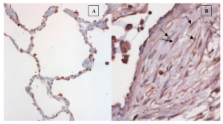
          Upregulation of AGTR1 occurred through the MKK1/MKK2 signalling pathway. Immunohistochemical staining showed AGTR1 expression by lung fibroblasts in fibroblastic foci within biopsies of idiopathic pulmonary fibrosis.

          Conclusions

          This study identifies several novel TGFβ targets in lung fibroblasts, and confirms with independent methods the induction of angiotensin II receptor type 1, underlining a potential role for angiotensin II receptor 1 antagonism in the treatment of lung fibrosis.

          Related collections

          Most cited references40

          • Record: found
          • Abstract: found
          • Article: not found

          Idiopathic pulmonary fibrosis: prevailing and evolving hypotheses about its pathogenesis and implications for therapy.

          Idiopathic pulmonary fibrosis is a progressive and usually fatal lung disease characterized by fibroblast proliferation and extracellular matrix remodeling, which result in irreversible distortion of the lung's architecture. Although the pathogenetic mechanisms remain to be determined, the prevailing hypothesis holds that fibrosis is preceded and provoked by a chronic inflammatory process that injures the lung and modulates lung fibrogenesis, leading to the end-stage fibrotic scar. However, there is little evidence that inflammation is prominent in early disease, and it is unclear whether inflammation is relevant to the development of the fibrotic process. Evidence suggests that inflammation does not play a pivotal role. Inflammation is not a prominent histopathologic finding, and epithelial injury in the absence of ongoing inflammation is sufficient to stimulate the development of fibrosis. In addition, the inflammatory response to a lung fibrogenic insult is not necessarily related to the fibrotic response. Clinical measurements of inflammation fail to correlate with stage or outcome, and potent anti-inflammatory therapy does not improve outcome. This review presents a growing body of evidence suggesting that idiopathic pulmonary fibrosis involves abnormal wound healing in response to multiple, microscopic sites of ongoing alveolar epithelial injury and activation associated with the formation of patchy fibroblast-myofibroblast foci, which evolve to fibrosis. Progress in understanding the fibrogenic mechanisms in the lung is likely to yield more effective therapies.
            Bookmark
            • Record: found
            • Abstract: found
            • Article: not found

            Adenovector-mediated gene transfer of active transforming growth factor-beta1 induces prolonged severe fibrosis in rat lung.

            Transforming growth factor (TGF)-beta1 has been implicated in the pathogenesis of fibrosis based upon its matrix-inducing effects on stromal cells in vitro, and studies demonstrating increased expression of total TGF-beta1 in fibrotic tissues from a variety of organs. The precise role in vivo of this cytokine in both its latent and active forms, however, remains unclear. Using replication-deficient adenovirus vectors to transfer the cDNA of porcine TGF-beta1 to rat lung, we have been able to study the effect of TGF-beta1 protein in the respiratory tract directly. We have demonstrated that transient overexpression of active, but not latent, TGF-beta1 resulted in prolonged and severe interstitial and pleural fibrosis characterized by extensive deposition of the extracellular matrix (ECM) proteins collagen, fibronectin, and elastin, and by emergence of cells with the myofibroblast phenotype. These results illustrate the role of TGF-beta1 and the importance of its activation in the pulmonary fibrotic process, and suggest that targeting active TGF-beta1 and steps involved in TGF-beta1 activation are likely to be valuable antifibrogenic therapeutic strategies. This new and versatile model of pulmonary fibrosis can be used to study such therapies.
              Bookmark
              • Record: found
              • Abstract: found
              • Article: not found

              PD 098059 is a specific inhibitor of the activation of mitogen-activated protein kinase kinase in vitro and in vivo.

              PD 098059 has been shown previously to inhibit the dephosphorylated form of mitogen-activated protein kinase kinase-1 (MAPKK1) and a mutant MAPKK1(S217E,S221E), which has low levels of constitutive activity (Dudley, D. T., Pang, L., Decker, S. J., Bridges, A. J., and Saltiel, A. R. (1995) Proc. Natl. Acad. Sci. U.S.A. 92, 7686-7689). Here we report that PD 098059 does not inhibit Raf-activated MAPKK1 but that it prevents the activation of MAPKK1 by Raf or MEK kinase in vitro at concentrations (IC50 = 2-7 microM) similar to those concentrations that inhibit dephosphorylated MAPKK1 or MAPKK1(S217E,S221E). PD 098059 inhibited the activation of MAPKK2 by Raf with a much higher IC50 value (50 microM) and did not inhibit the phosphorylation of other Raf or MEK kinase substrates, indicating that it exerts its effect by binding to the inactive form of MAPKK1. PD 098059 also acts as a specific inhibitor of the activation of MAPKK in Swiss 3T3 cells, suppressing by 80-90% its activation by a variety of agonists. The high degree of specificity of PD 098059 in vitro and in vivo is indicated by its failure to inhibit 18 protein Ser/Thr kinases (including two other MAPKK homologues) in vitro by its failure to inhibit the in vivo activation of MAPKK and MAP kinase homologues that participate in stress and interleukin-1-stimulated kinase cascades in KB and PC12 cells, and by lack of inhibition of the activation of p70 S6 kinase by insulin or epidermal growth factor in Swiss 3T3 cells. PD 098059 (50 microM) inhibited the activation of p42MAPK and isoforms of MAP kinase-activated protein kinase-1 in Swiss 3T3 cells, but the extent of inhibition depended on how potently c-Raf and MAPKK were activated by any particular agonist and demonstrated the enormous amplification potential of this kinase cascade. PD 098059 not only failed to inhibit the activation of Raf by platelet-derived growth factor, serum, insulin, and phorbol esters in Swiss 3T3 cells but actually enhanced Raf activity. The rate of activation of Raf by platelet-derived growth factor was increased 3-fold, and the subsequent inactivation that occurred after 10 min was prevented. These results indicate that the activation of Raf is suppressed and that its inactivation is accelerated by a downstream component(s) of the MAP kinase pathway.
                Bookmark

                Author and article information

                Journal
                Respir Res
                Respiratory Research
                BioMed Central
                1465-9921
                1465-993X
                2004
                30 November 2004
                : 5
                : 1
                : 24
                Affiliations
                [1 ]Interstitial Lung Disease Unit, Royal Brompton Hospital, Imperial College of Science, Technology and Medicine, Emmanuel Kaye Building, 1B Manresa Road, SW3 6LR, London, UK
                [2 ]Division of Academic Rheumatology, Royal Free Hospital, London, U.K
                [3 ]Centre for Cardiovascular Biology and Medicine, Guy's, King's, and St. Thomas' School of Biomedical Sciences, King's College London, UK
                [4 ]Division of Respiratory Diseases, University of Siena, Siena, Italy
                [5 ]MRC Clinical Science Centre, Hammersmith Campus, Imperial College London, UK
                [6 ]Dept of Pathology, Royal Brompton Hospital, London, UK
                Article
                1465-9921-5-24
                10.1186/1465-9921-5-24
                538264
                15571627
                0577246e-6ba8-492d-b68b-1be557c2ddad
                Copyright © 2004 Renzoni et al; licensee BioMed Central Ltd.

                This is an Open Access article distributed under the terms of the Creative Commons Attribution License ( http://creativecommons.org/licenses/by/2.0), which permits unrestricted use, distribution, and reproduction in any medium, provided the original work is properly cited.

                History
                : 5 September 2004
                : 30 November 2004
                Categories
                Research

                Respiratory medicine
                Respiratory medicine

                Comments

                Comment on this article