63
views
0
recommends
+1 Recommend
0 collections
    0
    shares
      • Record: found
      • Abstract: found
      • Article: found
      Is Open Access

      F-18 labelled PSMA-1007: biodistribution, radiation dosimetry and histopathological validation of tumor lesions in prostate cancer patients

      research-article

      Read this article at

          There is no author summary for this article yet. Authors can add summaries to their articles on ScienceOpen to make them more accessible to a non-specialist audience.

          Abstract

          Purpose

          The prostate-specific membrane antigen (PSMA) targeted positron-emitting-tomography (PET) tracer 68Ga-PSMA-11 shows great promise in the detection of prostate cancer. However, 68Ga has several shortcomings as a radiolabel including short half-life and non-ideal energies, and this has motivated consideration of 18F-labelled analogs. 18F-PSMA-1007 was selected among several 18F-PSMA-ligand candidate compounds because it demonstrated high labelling yields, outstanding tumor uptake and fast, non-urinary background clearance. Here, we describe the properties of 18F-PSMA-1007 in human volunteers and patients.

          Methods

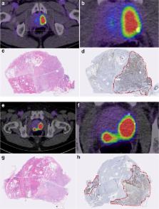
          Radiation dosimetry of 18F-PSMA-1007 was determined in three healthy volunteers who underwent whole-body PET-scans and concomitant blood and urine sampling. Following this, ten patients with high-risk prostate cancer underwent 18F-PSMA-1007 PET/CT (1 h and 3 h p.i.) and normal organ biodistribution and tumor uptakes were examined. Eight patients underwent prostatectomy with extended pelvic lymphadenectomy. Uptake in intra-prostatic lesions and lymph node metastases were correlated with final histopathology, including PSMA immunostaining.

          Results

          With an effective dose of approximately 4.4–5.5 mSv per 200–250 MBq examination, 18F-PSMA-1007 behaves similar to other PSMA-PET agents as well as to other 18F-labelled PET-tracers. In comparison to other PSMA-targeting PET-tracers, 18F-PSMA-1007 has reduced urinary clearance enabling excellent assessment of the prostate. Similar to 18F-DCFPyL and with slightly slower clearance kinetics than PSMA-11, favorable tumor-to-background ratios are observed 2–3 h after injection. In eight patients, diagnostic findings were successfully validated by histopathology. 18F-PSMA-1007 PET/CT detected 18 of 19 lymph node metastases in the pelvis, including nodes as small as 1 mm in diameter.

          Conclusion

          18F-PSMA-1007 performs at least comparably to 68Ga-PSMA-11, but its longer half-life combined with its superior energy characteristics and non-urinary excretion overcomes some practical limitations of 68Ga-labelled PSMA-targeted tracers.

          Electronic supplementary material

          The online version of this article (doi:10.1007/s00259-016-3573-4) contains supplementary material, which is available to authorized users.

          Related collections

          Most cited references11

          • Record: found
          • Abstract: found
          • Article: not found

          Initial Evaluation of [(18)F]DCFPyL for Prostate-Specific Membrane Antigen (PSMA)-Targeted PET Imaging of Prostate Cancer.

          Prostate-specific membrane antigen (PSMA) is a recognized target for imaging prostate cancer. Here we present initial safety, biodistribution, and radiation dosimetry results with [(18)F]DCFPyL, a second-generation fluorine-18-labeled small-molecule PSMA inhibitor, in patients with prostate cancer.
            Bookmark
            • Record: found
            • Abstract: found
            • Article: not found

            Biodistribution, tumor detection, and radiation dosimetry of 18F-DCFBC, a low-molecular-weight inhibitor of prostate-specific membrane antigen, in patients with metastatic prostate cancer.

            Prostate-specific membrane antigen (PSMA) is a type II integral membrane protein expressed on the surface of prostate cancer (PCa) cells, particularly in androgen-independent, advanced, and metastatic disease. Previously, we demonstrated that N-[N-[(S)-1,3-dicarboxypropyl]carbamoyl]-4-(18)F-fluorobenzyl-L-cysteine ((18)F-DCFBC) could image an experimental model of PSMA-positive PCa using PET. Here, we describe the initial clinical experience and radiation dosimetry of (18)F-DCFBC in men with metastatic PCa. Five patients with radiologic evidence of metastatic PCa were studied after the intravenous administration of 370 MBq (10 mCi) of (18)F-DCFBC. Serial PET was performed until 2 h after administration. Time-activity curves were generated for selected normal tissues and metastatic foci. Radiation dose estimates were calculated using OLINDA/EXM 1.1. Most vascular organs demonstrated a slow decrease in radioactivity concentration over time consistent with clearance from the blood pool, with primarily urinary radiotracer excretion. Thirty-two PET-positive suspected metastatic sites were identified, with 21 concordant on both PET and conventional imaging for abnormal findings compatible with metastatic disease. Of the 11 PET-positive sites not identified on conventional imaging, most were within the bone and could be considered suggestive for the detection of early bone metastases, although further validation is needed. The highest mean absorbed dose per unit administered radioactivity (μGy/MBq) was in the bladder wall (32.4), and the resultant effective dose was 19.9 ± 1.34 μSv/MBq (mean ± SD). Although further studies are needed for validation, our findings demonstrate the potential of (18)F-DCFBC as a new positron-emitting imaging agent for the detection of metastatic PCa. This study also provides dose estimates for (18)F-DCFBC that are comparable to those of other PET radiopharmaceuticals such as (18)F-FDG.
              Bookmark
              • Record: found
              • Abstract: found
              • Article: not found

              68Ga-PSMA-11 PET/CT: a new technique with high potential for the radiotherapeutic management of prostate cancer patients

              Purpose Radiotherapy is the main therapeutic approach besides surgery of localized prostate cancer. It relies on risk stratification and exact staging. This report analyses the potential of [68Ga]Glu-urea-Lys(Ahx)-HBED-CC (68Ga-PSMA-11), a new positron emission tomography (PET) tracer targeting prostate-specific membrane antigen (PSMA) for prostate cancer staging and individualized radiotherapy planning. Methods A cohort of 57 patients with prostate cancer scanned with 68Ga-PSMA-11 PET/CT for radiotherapy planning was retrospectively reviewed; 15 patients were at initial diagnosis and 42 patients at time of biochemical recurrence. Staging results of conventional imaging, including bone scintigraphy, CT or MRI, were compared with 68Ga-PSMA ligand PET/CT results and the influence on radiotherapeutic management was quantified. Results 68Ga-PSMA ligand PET/CT had a dramatic impact on radiotherapy application in the presented cohort. In 50.8 % of the cases therapy was changed. Conclusion The presented imaging technique of 68Ga-PSMA PET/CT could be a key technology for individualized radiotherapy management in prostate cancer.
                Bookmark

                Author and article information

                Contributors
                +49-6221-56-7732 , frederik@egiesel.com
                Journal
                Eur J Nucl Med Mol Imaging
                Eur. J. Nucl. Med. Mol. Imaging
                European Journal of Nuclear Medicine and Molecular Imaging
                Springer Berlin Heidelberg (Berlin/Heidelberg )
                1619-7070
                1619-7089
                26 November 2016
                26 November 2016
                2017
                : 44
                : 4
                : 678-688
                Affiliations
                [1 ]ISNI 0000 0001 0328 4908, GRID grid.5253.1, Department of Nuclear Medicine, , University Hospital Heidelberg, ; INF 400, 69120 Heidelberg, Germany
                [2 ]ISNI 0000 0001 0328 4908, GRID grid.5253.1, Department of Urology, , University Hospital Heidelberg, ; Heidelberg, Germany
                [3 ]ISNI 0000 0004 0492 0584, GRID grid.7497.d, Division of Radiopharmaceutical Chemistry, , German Cancer Research Center (dkfz), ; Heidelberg, Germany
                [4 ]ABX-CRO, Dresden, Germany
                [5 ]ISNI 0000 0001 0328 4908, GRID grid.5253.1, Section of Molecular Urooncology, Department of Urology, Medical Faculty Heidelberg, , University Hospital Heidelberg, ; Heidelberg, Germany
                [6 ]ISNI 0000 0001 0328 4908, GRID grid.5253.1, Institute of Pathology, , University Hospital Heidelberg, ; Heidelberg, Germany
                [7 ]ISNI 0000 0001 0328 4908, GRID grid.5253.1, Department of Medical Oncology, National Center for Tumor Diseases (NCT), , University Hospital Heidelberg, ; Heidelberg, Germany
                [8 ]ISNI 0000 0001 2190 4373, GRID grid.7700.0, Hamamatsu Tissue Imaging and Analysis Center, , University of Heidelberg, ; Heidelberg, Germany
                Article
                3573
                10.1007/s00259-016-3573-4
                5323462
                27889802
                92c33f85-e7db-4f05-b2b6-1af50ec2dc87
                © The Author(s) 2016

                Open Access This article is distributed under the terms of the Creative Commons Attribution 4.0 International License (http://creativecommons.org/licenses/by/4.0/), which permits unrestricted use, distribution, and reproduction in any medium, provided you give appropriate credit to the original author(s) and the source, provide a link to the Creative Commons license, and indicate if changes were made.

                History
                : 2 August 2016
                : 9 November 2016
                Categories
                Original Article
                Custom metadata
                © Springer-Verlag Berlin Heidelberg 2017

                Radiology & Imaging
                18f-psma,f-18-psma,psma-1007,pet/ct,positron emission tomography
                Radiology & Imaging
                18f-psma, f-18-psma, psma-1007, pet/ct, positron emission tomography

                Comments

                Comment on this article