122
views
0
recommends
+1 Recommend
0 collections
    0
    shares
      • Record: found
      • Abstract: found
      • Article: not found

      Coordination of Rho GTPase activities during cell protrusion

      research-article

      Read this article at

      ScienceOpenPublisherPMC
          There is no author summary for this article yet. Authors can add summaries to their articles on ScienceOpen to make them more accessible to a non-specialist audience.

          Abstract

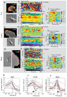
          The GTPases Rac1, RhoA and Cdc42 act in concert to control cytoskeleton dynamics 1- 3. Recent biosensor studies have shown that all three GTPases are activated at the front of migrating cells 4- 7 and biochemical evidence suggests that they may regulate one another: Cdc42 can activate Rac1 8, and Rac1 and RhoA are mutually inhibitory 9- 12. However, their spatiotemporal coordination, at the seconds and single micron dimensions typical of individual protrusion events, remains unknown. Here, we examine GTPase coordination both through simultaneous visualization of two GTPase biosensors and using a “computational multiplexing” approach capable of defining the relationships between multiple protein activities visualized in separate experiments. We found that RhoA is activated at the cell edge synchronous with edge advancement, whereas Cdc42 and Rac1 are activated 2 μm behind the edge with a delay of 40 sec. This indicates that Rac1 and RhoA operate antagonistically through spatial separation and precise timing, and that RhoA plays a role in the initial events of protrusion, while Rac1 and Cdc42 activate pathways implicated in reinforcement and stabilization of newly expanded protrusions.

          Related collections

          Most cited references20

          • Record: found
          • Abstract: found
          • Article: not found

          Localized Rac activation dynamics visualized in living cells.

          Signaling proteins are thought to be tightly regulated spatially and temporally in order to generate specific and localized effects. For Rac and other small guanosine triphosphatases, binding to guanosine triphosphate leads to interaction with downstream targets and regulates subcellular localization. A method called FLAIR (fluorescence activation indicator for Rho proteins) was developed to quantify the spatio-temporal dynamics of the Rac1 nucleotide state in living cells. FLAIR revealed precise spatial control of growth factor-induced Rac activation, in membrane ruffles and in a gradient of activation at the leading edge of motile cells. FLAIR exemplifies a generally applicable approach for examining spatio-temporal control of protein activity.
            Bookmark
            • Record: found
            • Abstract: found
            • Article: not found

            Interplay between Rac and Rho in the control of substrate contact dynamics.

            Substrate anchorage and cell locomotion entail the initiation and development of different classes of contact sites, which are associated with the different compartments of the actin cytoskeleton. The Rho-family GTPases are implicated in the signalling pathways that dictate contact initiation, maturation and turnover, but their individual roles in these processes remain to be defined. We monitored the dynamics of peripheral, Rac-induced focal complexes in living cells in response to perturbations of Rac and Rho activity and myosin contractility. We show that focal complexes formed in response to Rac differentiated into focal contacts upon upregulation of Rho. Focal complexes were dissociated by inhibitors of myosin-II-dependent contractility but not by an inhibitor of Rho-kinase. The downregulation of Rac promoted the enlargement of focal contacts, whereas a block in the Rho pathway not only caused a dissolution of focal contacts but also stimulated membrane ruffling and formation of new focal complexes, which were associated with the advance of the cell front. Rac functions to signal the creation of new substrate contacts at the cell front, which are associated with the induction of ruffling lamellipodia, whereas Rho serves in the maturation of existing contacts, with both contact types requiring contractility for their formation. The transition from a focal complex to a focal contact is associated with a switch to Rho-kinase dependence. Rac and Rho also influence the development of focal contacts and focal complexes, respectively, through mutually antagonistic pathways.
              Bookmark
              • Record: found
              • Abstract: found
              • Article: not found

              Paxillin phosphorylation at Ser273 localizes a GIT1–PIX–PAK complex and regulates adhesion and protrusion dynamics

              Continuous adhesion formation and disassembly (adhesion turnover) in the protrusions of migrating cells is regulated by unclear mechanisms. We show that p21-activated kinase (PAK)–induced phosphorylation of serine 273 in paxillin is a critical regulator of this turnover. Paxillin-S273 phosphorylation dramatically increases migration, protrusion, and adhesion turnover by increasing paxillin–GIT1 binding and promoting the localization of a GIT1–PIX–PAK signaling module near the leading edge. Mutants that interfere with the formation of this ternary module abrogate the effects of paxillin-S273 phosphorylation. PAK-dependent paxillin-S273 phosphorylation functions in a positive-feedback loop, as active PAK, active Rac, and myosin II activity are all downstream effectors of this turnover pathway. Finally, our studies led us to identify in highly motile cells a class of small adhesions that reside near the leading edge, turnover in 20–30 s, and resemble those seen with paxillin-S273 phosphorylation. These adhesions appear to be regulated by the GIT1–PIX–PAK module near the leading edge.
                Bookmark

                Author and article information

                Journal
                0410462
                6011
                Nature
                Nature
                0028-0836
                1476-4687
                27 April 2010
                19 August 2009
                3 September 2009
                14 June 2010
                : 461
                : 7260
                : 99-103
                Affiliations
                [1 ]Department of Cell Biology, The Scripps Research Institute, 10550 N. Torrey Pines Road, La Jolla, CA 92037 USA
                [2 ]Departments of Pharmacology, Medicinal Chemistry and Lineberger Cancer Center, University of North Carolina at Chapel Hill, Chapel Hill, NC 27599 USA
                Author notes
                Author Information Correspondence and requests for materials should be addressed to K.M.H. ( khahn@ 123456med.unc.edu ) or G.D. ( gdanuser@ 123456scripps.edu )
                [3]

                Current address: Novartis Pharma AG, Lichtstrasse 35, CH-4056 Basel, Switzerland

                [4]

                Current address: Department of Anatomy and Structural Biology and Gruss-Lipper Biophotonics Center, Albert Einstein College of Medicine of Yeshiva University, 1300 Morris Park Ave, Bronx, NY 10461 USA

                [5]

                Current address: Department of Biomedicine, University of Basel, Mattenstrasse 28, CH-4058 Basel, Switzerland

                [6]

                Current address: Department of Molecular Cell Biology, University of Duisburg-Essen, Universitaetsstrasse 2, 45117 Essen, Germany

                [*]

                These authors contributed equally to this work.

                Article
                nihpa127665
                10.1038/nature08242
                2885353
                19693013
                31bb29b6-5e30-40a0-89b7-143dac020b07
                History
                Funding
                Funded by: National Institute of General Medical Sciences : NIGMS
                Award ID: U54 GM064346-099029 ||GM
                Funded by: National Institute of General Medical Sciences : NIGMS
                Award ID: R01 GM071868-04 ||GM
                Funded by: National Institute of General Medical Sciences : NIGMS
                Award ID: R01 GM057464-09 ||GM
                Categories
                Article

                Uncategorized
                Uncategorized

                Comments

                Comment on this article