12
views
0
recommends
+1 Recommend
0 collections
    0
    shares
      • Record: found
      • Abstract: found
      • Article: found
      Is Open Access

      Silver Nanoparticles in the Lung: Toxic Effects and Focal Accumulation of Silver in Remote Organs

      research-article

      Read this article at

          There is no author summary for this article yet. Authors can add summaries to their articles on ScienceOpen to make them more accessible to a non-specialist audience.

          Abstract

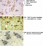
          The distribution of silver (Ag) into remote organs secondary to the application of Ag nanoparticles (Ag-NP) to the lung is still incompletely understood and was investigated in the rat with imaging methods. Dose-finding experiments were carried out with 50 nm- or 200 nm-sized polyvinyl pyrrolidine (PVP)-coated Ag-NP using alveolar macrophages in vitro and female rats, which received Ag-NP via intratracheal instillation. In the main study, we administered 37.5–300 µg per rat lung of the more toxic Ag50-PVP and assessed the broncho-alveolar lavage fluid (BALF) for inflammatory cells, total protein and fibronectin after three and 21 days. In parallel, lung tissue was analysed for DNA double-strand breaks and altered cell proliferation. While 75–150 µg Ag50-PVP per rat lung caused a reversible inflammation, 300 µg led to DNA damage, accelerated cell proliferation and progressively increasing numbers of neutrophilic granulocytes. Ag accumulation was significant in homogenates of liver and other peripheral organs upon lung dose of ≥75 µg. Quantitative laser-ablation inductively-coupled plasma mass spectrometry (LA-ICP-MS) combined with enhanced dark field microscopy and autometallography revealed focal accumulations of Ag and/or Ag-NP in sections of peripheral organs: mediastinal lymph nodes contained Ag-NP especially in peripheral macrophages and Ag in argyrophilic fibres. In the kidney, Ag had accumulated within proximal tubuli, while renal filter structures contained no Ag. Discrete localizations were also observed in immune cells of liver and spleen. Overall, the study shows that concentrations of Ag-NP, which elicit a transient inflammation in the rat lung, lead to focal accumulations of Ag in peripheral organs, and this might pose a risk to particular cell populations in remote sites.

          Related collections

          Most cited references54

          • Record: found
          • Abstract: found
          • Article: found
          Is Open Access

          Size-dependent cytotoxicity of silver nanoparticles in human lung cells: the role of cellular uptake, agglomeration and Ag release

          Background Silver nanoparticles (AgNPs) are currently one of the most manufactured nanomaterials. A wide range of toxicity studies have been performed on various AgNPs, but these studies report a high variation in toxicity and often lack proper particle characterization. The aim of this study was to investigate size- and coating-dependent toxicity of thoroughly characterized AgNPs following exposure of human lung cells and to explore the mechanisms of toxicity. Methods BEAS-2B cells were exposed to citrate coated AgNPs of different primary particle sizes (10, 40 and 75 nm) as well as to 10 nm PVP coated and 50 nm uncoated AgNPs. The particle agglomeration in cell medium was investigated by photon cross correlation spectroscopy (PCCS); cell viability by LDH and Alamar Blue assay; ROS induction by DCFH-DA assay; genotoxicity by alkaline comet assay and γH2AX foci formation; uptake and intracellular localization by transmission electron microscopy (TEM); and cellular dose as well as Ag release by atomic absorption spectroscopy (AAS). Results The results showed cytotoxicity only of the 10 nm particles independent of surface coating. In contrast, all AgNPs tested caused an increase in overall DNA damage after 24 h assessed by the comet assay, suggesting independent mechanisms for cytotoxicity and DNA damage. However, there was no γH2AX foci formation and no increased production of intracellular reactive oxygen species (ROS). The reasons for the higher toxicity of the 10 nm particles were explored by investigating particle agglomeration in cell medium, cellular uptake, intracellular localization and Ag release. Despite different agglomeration patterns, there was no evident difference in the uptake or intracellular localization of the citrate and PVP coated AgNPs. However, the 10 nm particles released significantly more Ag compared with all other AgNPs (approx. 24 wt% vs. 4–7 wt%) following 24 h in cell medium. The released fraction in cell medium did not induce any cytotoxicity, thus implying that intracellular Ag release was responsible for the toxicity. Conclusions This study shows that small AgNPs (10 nm) are cytotoxic for human lung cells and that the toxicity observed is associated with the rate of intracellular Ag release, a ‘Trojan horse’ effect.
            Bookmark
            • Record: found
            • Abstract: found
            • Article: found
            Is Open Access

            Tissue macrophages: heterogeneity and functions

            Macrophages are present in all vertebrate tissues, from mid-gestation throughout life, constituting a widely dispersed organ system. They promote homeostasis by responding to internal and external changes within the body, not only as phagocytes in defence against microbes and in clearance of dead and senescent cells, but also through trophic, regulatory and repair functions. In this review, we describe macrophage phenotypic heterogeneity in different tissue environments, drawing particular attention to organ-specific functions. Electronic supplementary material The online version of this article (doi:10.1186/s12915-017-0392-4) contains supplementary material, which is available to authorized users.
              Bookmark
              • Record: found
              • Abstract: found
              • Article: not found

              Oxidative stress-dependent toxicity of silver nanoparticles in human hepatoma cells.

              Cytotoxicity induced by silver nanoparticles (AgNPs) and the role that oxidative stress plays in this process were demonstrated in human hepatoma cells. Toxicity induced by silver (Ag(+)) ions was studied in parallel using AgNO(3) as the Ag(+) ion source. Using cation exchange treatment, we confirmed that the AgNP solution contained a negligible amount of free Ag(+) ions. Metal-responsive metallothionein 1b (MT1b) mRNA expression was not induced in AgNP-treated cells, while it was induced in AgNO(3)-treated cells. These results indicate that AgNP-treated cells have limited exposure to Ag(+) ions, despite the potential release of Ag(+) ions from AgNPs in cell culture. AgNPs agglomerated in the cytoplasm and nuclei of treated cells, and induced intracellular oxidative stress. AgNPs exhibited cytotoxicity with a potency comparable to that of Ag(+) ions in in vitro cytotoxicity assays. However, the toxicity of AgNPs was prevented by use of the antioxidant N-acetylcysteine, and AgNP-induced DNA damage was also prevented by N-acetylcysteine. AgNO(3) treatment induced oxidative stress-related glutathione peroxidase 1 (GPx1) and catalase expression to a greater extent than AgNP exposure, but treatment with AgNO(3) and AgNPs induced comparable superoxide dismutase 1 (SOD1) expression levels. Our findings suggest that AgNP cytotoxicity is primarily the result of oxidative stress and is independent of the toxicity of Ag(+) ions.
                Bookmark

                Author and article information

                Journal
                Nanomaterials (Basel)
                Nanomaterials (Basel)
                nanomaterials
                Nanomaterials
                MDPI
                2079-4991
                12 December 2017
                December 2017
                : 7
                : 12
                : 441
                Affiliations
                [1 ]IBE R&D Institute for Lung Health gGmbH, Mendelstr. 11, 48149 Münster, Germany; vennemann@ 123456ibe-ms.de
                [2 ]Institute of Inorganic and Analytical Chemistry, University of Münster, Corrensstr. 30, 48149 Münster, Germany; franziska.blaske@ 123456googlemail.com (F.B.); msperlin@ 123456uni-muenster.de (M.S.); uk@ 123456uni-muenster.de (U.K.)
                Author notes
                [* ]Correspondence: martin.wiemann@ 123456ibe-ms.de ; Tel.: +49-251-9802340
                Author information
                https://orcid.org/0000-0001-6676-2487
                https://orcid.org/0000-0002-1774-6787
                Article
                nanomaterials-07-00441
                10.3390/nano7120441
                5746931
                29231883
                516feec5-4990-47f6-8bc9-0651d0dd87aa
                © 2017 by the authors.

                Licensee MDPI, Basel, Switzerland. This article is an open access article distributed under the terms and conditions of the Creative Commons Attribution (CC BY) license ( http://creativecommons.org/licenses/by/4.0/).

                History
                : 30 October 2017
                : 05 December 2017
                Categories
                Article

                silver nanoparticle,quantitative bio-imaging,in vitro toxicity,nanoparticle transition

                Comments

                Comment on this article