79
views
0
recommends
+1 Recommend
0 collections
    0
    shares
      • Record: found
      • Abstract: found
      • Article: not found

      Block of A1 astrocyte conversion by microglia is neuroprotective in models of Parkinson’s disease

      research-article
      1 , 2 , 15 , 1 , 2 , 1 , 2 , 1 , 2 , 6 , 7 , 6 , 7 , 1 , 2 , 6 , 7 , 1 , 2 , 15 , 1 , 2 , 1 , 2 , 1 , 2 , 1 , 2 , 1 , 2 , 1 , 2 , 15 , 1 , 2 , 15 , 8 , 1 , 2 , 1 , 2 , 1 , 2 , 9 , 10 , 11 , 1 , 2 , 3 , 4 , 12 , 13 , 14 , 2 , 14 , 1 , 2 , 3 , 4 , 15 , 6 , 7 , 1 , 2 , 4 , 5 , 15 , 16 , 1 , 2 , 15 , 16
      Nature medicine

      Read this article at

      ScienceOpenPublisherPMC
          There is no author summary for this article yet. Authors can add summaries to their articles on ScienceOpen to make them more accessible to a non-specialist audience.

          Abstract

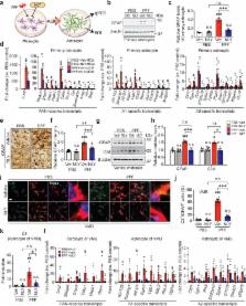
          Activation of microglia by classical inflammatory mediators can convert astrocytes to a neurotoxic A1 phenotype in a variety of neurological diseases 1, 2 . Development of agents that could inhibit the formation of A1 reactive astrocytes could be used to treat these diseases for which there are no disease modifying therapies. Glucagon-like peptide-1 receptor (GLP-1R) agonists have been touted as potential neuroprotective agents for neurologic disorders such as Alzheimer’s disease (AD) and Parkinson’s disease (PD) 3- 13 . The mechanisms by which GLP-1R agonists are neuroprotective are not known. Here we show that a potent, brain penetrant long acting GLP-1R agonist NLY01 protects against the loss of dopamine neurons and behavioral deficits in the α-synuclein preformed fibril (α-syn PFF) model of sporadic PD 14, 15 . NLY01 also prolongs the life and reduces the behavioral deficits and neuropathological abnormalities in the human A53T α-synuclein (hA53T) transgenic (Tg) model of α-synucleinopathy induced neurodegeneration 16 . We found that NLY01 is a potent GLP-1R agonist with favorable properties that is neuroprotective via the direct prevention of microglial mediated conversion of astrocytes to an A1 neurotoxic phenotype. In light of NLY01 favorable properties it should be evaluated in the treatment of PD and related neurologic disorders characterized by microglial activation.

          Related collections

          Most cited references32

          • Record: found
          • Abstract: found
          • Article: not found

          GLP-1 receptor agonists for individualized treatment of type 2 diabetes mellitus.

          In healthy humans, the incretin glucagon-like peptide 1 (GLP-1) is secreted after eating and lowers glucose concentrations by augmenting insulin secretion and suppressing glucagon release. Additional effects of GLP-1 include retardation of gastric emptying, suppression of appetite and, potentially, inhibition of β-cell apoptosis. Native GLP-1 is degraded within ~2-3 min in the circulation; various GLP-1 receptor agonists have, therefore, been developed to provide prolonged in vivo actions. These GLP-1 receptor agonists can be categorized as either short-acting compounds, which provide short-lived receptor activation (such as exenatide and lixisenatide) or as long-acting compounds (for example albiglutide, dulaglutide, exenatide long-acting release, and liraglutide), which activate the GLP-1 receptor continuously at their recommended dose. The pharmacokinetic differences between these drugs lead to important differences in their pharmacodynamic profiles. The short-acting GLP-1 receptor agonists primarily lower postprandial blood glucose levels through inhibition of gastric emptying, whereas the long-acting compounds have a stronger effect on fasting glucose levels, which is mediated predominantly through their insulinotropic and glucagonostatic actions. The adverse effect profiles of these compounds also differ. The individual properties of the various GLP-1 receptor agonists might enable incretin-based treatment of type 2 diabetes mellitus to be tailored to the needs of each patient.
            Bookmark
            • Record: found
            • Abstract: found
            • Article: not found

            Addition of exogenous α-synuclein preformed fibrils to primary neuronal cultures to seed recruitment of endogenous α-synuclein to Lewy body and Lewy neurite-like aggregates.

            This protocol describes a primary neuronal model of formation of α-synuclein (α-syn) aggregates that recapitulate features of the Lewy bodies and Lewy neurites found in Parkinson's disease brains and other synucleinopathies. This model allows investigation of aggregate formation, their impact on neuron function, and development of therapeutics. Addition of preformed fibrils (PFFs) synthesized from recombinant α-syn to neurons seeds the recruitment of endogenous α-syn into aggregates characterized by detergent insolubility and hyperphosphorylation. Aggregate formation follows a lag phase of 2-3 d, followed by formation in axons by days 4-7, spread to somatodendritic compartments by days 7-10 and neuron death ~14 d after PFF addition. Here we provide methods and highlight the crucial steps for PFF formation, PFF addition to cultured hippocampal neurons and confirmation of aggregate formation. Neurons derived from various brain regions from nontransgenic and genetically engineered mice and rats can be used, allowing interrogation of the effect of specific genes on aggregate formation.
              Bookmark
              • Record: found
              • Abstract: found
              • Article: not found

              GLP-1 receptor stimulation preserves primary cortical and dopaminergic neurons in cellular and rodent models of stroke and Parkinsonism.

              Glucagon-like peptide-1 (GLP-1) is an endogenous insulinotropic peptide secreted from the gastrointestinal tract in response to food intake. It enhances pancreatic islet beta-cell proliferation and glucose-dependent insulin secretion, and lowers blood glucose and food intake in patients with type 2 diabetes mellitus (T2DM). A long-acting GLP-1 receptor (GLP-1R) agonist, exendin-4 (Ex-4), is the first of this new class of antihyperglycemia drugs approved to treat T2DM. GLP-1Rs are coupled to the cAMP second messenger pathway and, along with pancreatic cells, are expressed within the nervous system of rodents and humans, where receptor activation elicits neurotrophic actions. We detected GLP-1R mRNA expression in both cultured embryonic primary cerebral cortical and ventral mesencephalic (dopaminergic) neurons. These cells are vulnerable to hypoxia- and 6-hydroxydopamine-induced cell death, respectively. We found that GLP-1 and Ex-4 conferred protection in these cells, but not in cells from Glp1r knockout (-/-) mice. Administration of Ex-4 reduced brain damage and improved functional outcome in a transient middle cerebral artery occlusion stroke model. Ex-4 treatment also protected dopaminergic neurons against degeneration, preserved dopamine levels, and improved motor function in the 1-methyl-4-phenyl-1,2,3,6-tetrahydropyridine (MPTP) mouse model of Parkinson's disease (PD). Our findings demonstrate that Ex-4 can protect neurons against metabolic and oxidative insults, and they provide preclinical support for the therapeutic potential for Ex-4 in the treatment of stroke and PD.
                Bookmark

                Author and article information

                Journal
                9502015
                8791
                Nat Med
                Nat. Med.
                Nature medicine
                1078-8956
                1546-170X
                28 April 2018
                11 June 2018
                July 2018
                11 December 2018
                : 24
                : 7
                : 931-938
                Affiliations
                [1 ]Neuroregeneration and Stem Cell Programs, Institute for Cell Engineering, The Johns Hopkins University School of Medicine, Baltimore, MD 21205, USA
                [2 ]Department of Neurology, The Johns Hopkins University School of Medicine, Baltimore, MD 21205, USA
                [3 ]Department of Physiology, The Johns Hopkins University School of Medicine, Baltimore, MD 21205, USA
                [4 ]Solomon H. Snyder Department of Neuroscience, The Johns Hopkins University School of Medicine, Baltimore, MD 21205, USA
                [5 ]Department of Pharmacology and Molecular Sciences, The Johns Hopkins University School of Medicine, Baltimore, MD 21205, USA
                [6 ]The Russell H. Morgan Department of Radiology and Radiological Sciences, Johns Hopkins University School of Medicine, Baltimore, MD 21287, USA
                [7 ]The Center for Nanomedicine at the Wilmer Eye Institute, Johns Hopkins University School of Medicine, Baltimore, MD 21287, USA
                [8 ]Department of Pharmacology and Toxicology, University of Alabama at Birmingham School of Medicine, Birmingham, AL 35294, USA
                [9 ]Division of Pharmacology, Department of Molecular Cell Biology, Sungkyunkwan University School of Medicine, Samsung Biomedical Research Institute, Suwon 16419, Republic of Korea
                [10 ]College of Pharmacy, Sungkyunkwan University, Suwon 16419, Republic of Korea
                [11 ]College of Pharmacy, Chung-Ang University, Seoul 06974, Republic of Korea
                [12 ]Soonchunhyang Medical Science Research Institute, Soonchunhyang University, Seoul Hospital, Seoul 04401, Republic of Korea
                [13 ]Neuraly Inc. Baltimore, MD 21205, USA
                [14 ]Department of Neurobiology, Stanford University, School of Medicine, Stanford, CA 94305, USA
                [15 ]Adrienne Helis Malvin Medical Research Foundation, New Orleans, LA 70130, USA
                [16 ]Diana Helis Henry Medical Research Foundation, New Orleans, LA 70130, USA
                Author notes
                Correspondence should be addressed to: Seulki Lee, Ph.D., The Russell H. Morgan Department of Radiology and Radiological Science, Center for Nanomedicine, Johns Hopkins University School of Medicine, 400 North Broadway, Baltimore, MD 21287, USA. slee343@ 123456jhmi.edu ; Ted M. Dawson, M.D., Ph.D., Neuroregeneration and Stem Cell Programs, Institute for Cell Engineering, Johns Hopkins University School of Medicine, 733 North Broadway, Baltimore, MD 21205, USA. tdawson@ 123456jhmi.edu ; Han Seok Ko, Ph.D., Neuroregeneration and Stem Cell Programs, Institute for Cell Engineering, Johns Hopkins University School of Medicine, 733 North Broadway, Baltimore, MD 21205, USA, hko3@ 123456jhmi.edu
                [*]

                These authors contributed equally.

                Article
                NIHMS958030
                10.1038/s41591-018-0051-5
                6039259
                29892066
                006dc8ff-e205-495b-b936-a00efa9723f9

                Users may view, print, copy, and download text and data-mine the content in such documents, for the purposes of academic research, subject always to the full Conditions of use: http://www.nature.com/authors/editorial_policies/license.html#terms

                History
                Categories
                Article

                Medicine
                Medicine

                Comments

                Comment on this article