0
views
0
recommends
+1 Recommend
0 collections
    0
    shares
      • Record: found
      • Abstract: found
      • Article: found
      Is Open Access

      Selection of pre-trained weights for transfer learning in automated cytomegalovirus retinitis classification

      research-article

      Read this article at

      Bookmark
          There is no author summary for this article yet. Authors can add summaries to their articles on ScienceOpen to make them more accessible to a non-specialist audience.

          Abstract

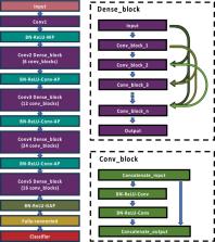
          Cytomegalovirus retinitis (CMVR) is a significant cause of vision loss. Regular screening is crucial but challenging in resource-limited settings. A convolutional neural network is a state-of-the-art deep learning technique to generate automatic diagnoses from retinal images. However, there are limited numbers of CMVR images to train the model properly. Transfer learning (TL) is a strategy to train a model with a scarce dataset. This study explores the efficacy of TL with different pre-trained weights for automated CMVR classification using retinal images. We utilised a dataset of 955 retinal images (524 CMVR and 431 normal) from Siriraj Hospital, Mahidol University, collected between 2005 and 2015. Images were processed using Kowa VX-10i or VX-20 fundus cameras and augmented for training. We employed DenseNet121 as a backbone model, comparing the performance of TL with weights pre-trained on ImageNet, APTOS2019, and CheXNet datasets. The models were evaluated based on accuracy, loss, and other performance metrics, with the depth of fine-tuning varied across different pre-trained weights. The study found that TL significantly enhances model performance in CMVR classification. The best results were achieved with weights sequentially transferred from ImageNet to APTOS2019 dataset before application to our CMVR dataset. This approach yielded the highest mean accuracy (0.99) and lowest mean loss (0.04), outperforming other methods. The class activation heatmaps provided insights into the model's decision-making process. The model with APTOS2019 pre-trained weights offered the best explanation and highlighted the pathologic lesions resembling human interpretation. Our findings demonstrate the potential of sequential TL in improving the accuracy and efficiency of CMVR diagnosis, particularly in settings with limited data availability. They highlight the importance of domain-specific pre-training in medical image classification. This approach streamlines the diagnostic process and paves the way for broader applications in automated medical image analysis, offering a scalable solution for early disease detection.

          Related collections

          Most cited references18

          • Record: found
          • Abstract: not found
          • Article: not found

          A Survey on Transfer Learning

            Bookmark
            • Record: found
            • Abstract: found
            • Article: found
            Is Open Access

            Artificial intelligence and deep learning in ophthalmology

            Artificial intelligence (AI) based on deep learning (DL) has sparked tremendous global interest in recent years. DL has been widely adopted in image recognition, speech recognition and natural language processing, but is only beginning to impact on healthcare. In ophthalmology, DL has been applied to fundus photographs, optical coherence tomography and visual fields, achieving robust classification performance in the detection of diabetic retinopathy and retinopathy of prematurity, the glaucoma-like disc, macular oedema and age-related macular degeneration. DL in ocular imaging may be used in conjunction with telemedicine as a possible solution to screen, diagnose and monitor major eye diseases for patients in primary care and community settings. Nonetheless, there are also potential challenges with DL application in ophthalmology, including clinical and technical challenges, explainability of the algorithm results, medicolegal issues, and physician and patient acceptance of the AI ‘black-box’ algorithms. DL could potentially revolutionise how ophthalmology is practised in the future. This review provides a summary of the state-of-the-art DL systems described for ophthalmic applications, potential challenges in clinical deployment and the path forward.
              Bookmark
              • Record: found
              • Abstract: not found
              • Article: not found

              Medical Image Analysis using Convolutional Neural Networks: A Review

                Bookmark

                Author and article information

                Contributors
                worapan.kun@mahidol.edu
                Journal
                Sci Rep
                Sci Rep
                Scientific Reports
                Nature Publishing Group UK (London )
                2045-2322
                10 July 2024
                10 July 2024
                2024
                : 14
                : 15899
                Affiliations
                [1 ]GRID grid.10223.32, ISNI 0000 0004 1937 0490, Department of Ophthalmology, Faculty of Medicine Siriraj Hospital, , Mahidol University, ; Bangkok, Thailand
                [2 ]Present Address: Faculty of Information and Communication Technology, Mahidol University, ( https://ror.org/01znkr924) Nakhon Pathom, Thailand
                Article
                67121
                10.1038/s41598-024-67121-7
                11237151
                38987446
                ff575be6-be19-46b0-8934-eabbbec7accf
                © The Author(s) 2024

                Open Access This article is licensed under a Creative Commons Attribution 4.0 International License, which permits use, sharing, adaptation, distribution and reproduction in any medium or format, as long as you give appropriate credit to the original author(s) and the source, provide a link to the Creative Commons licence, and indicate if changes were made. The images or other third party material in this article are included in the article's Creative Commons licence, unless indicated otherwise in a credit line to the material. If material is not included in the article's Creative Commons licence and your intended use is not permitted by statutory regulation or exceeds the permitted use, you will need to obtain permission directly from the copyright holder. To view a copy of this licence, visit http://creativecommons.org/licenses/by/4.0/.

                History
                : 27 January 2024
                : 8 July 2024
                Categories
                Article
                Custom metadata
                © Springer Nature Limited 2024

                Uncategorized
                machine learning,retinal diseases,viral infection,computer science
                Uncategorized
                machine learning, retinal diseases, viral infection, computer science

                Comments

                Comment on this article