3
views
0
recommends
+1 Recommend
0 collections
    0
    shares
      • Record: found
      • Abstract: found
      • Article: found
      Is Open Access

      Predicting breast cancer response to neoadjuvant treatment using multi-feature MRI: results from the I-SPY 2 TRIAL

      research-article
      1 , 1 , 1 , 1 , 1 , 1 , 1 , 2 , 1 , 1 , 1 , 1 , 1 , 3 , 3 , 4 , 4 , 4 , 4 , 5 , 5 , 5 , 6 , 6 , 6 , 7 , 7 , 7 , 7 , 8 , 9 , 9 , 9 , 10 , 10 , 11 , 11 , 12 , 12 , 13 , 13 , 14 , 14 , 14 , 15 , 15 , 16 , 16 , 17 , 18 , 19 , 19 , 20 , 20 , 21 , 21 , 22 , 23 , 1 , 9 , 24 , 1 , 1 ,
      NPJ Breast Cancer
      Nature Publishing Group UK
      Breast cancer, Cancer imaging

      Read this article at

      Bookmark
          There is no author summary for this article yet. Authors can add summaries to their articles on ScienceOpen to make them more accessible to a non-specialist audience.

          Abstract

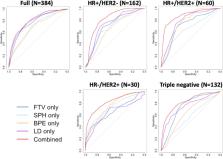
          Dynamic contrast-enhanced (DCE) MRI provides both morphological and functional information regarding breast tumor response to neoadjuvant chemotherapy (NAC). The purpose of this retrospective study is to test if prediction models combining multiple MRI features outperform models with single features. Four features were quantitatively calculated in each MRI exam: functional tumor volume, longest diameter, sphericity, and contralateral background parenchymal enhancement. Logistic regression analysis was used to study the relationship between MRI variables and pathologic complete response (pCR). Predictive performance was estimated using the area under the receiver operating characteristic curve (AUC). The full cohort was stratified by hormone receptor (HR) and human epidermal growth factor receptor 2 (HER2) status (positive or negative). A total of 384 patients (median age: 49 y/o) were included. Results showed analysis with combined features achieved higher AUCs than analysis with any feature alone. AUCs estimated for the combined versus highest AUCs among single features were 0.81 (95% confidence interval [CI]: 0.76, 0.86) versus 0.79 (95% CI: 0.73, 0.85) in the full cohort, 0.83 (95% CI: 0.77, 0.92) versus 0.73 (95% CI: 0.61, 0.84) in HR-positive/HER2-negative, 0.88 (95% CI: 0.79, 0.97) versus 0.78 (95% CI: 0.63, 0.89) in HR-positive/HER2-positive, 0.83 (95% CI not available) versus 0.75 (95% CI: 0.46, 0.81) in HR-negative/HER2-positive, and 0.82 (95% CI: 0.74, 0.91) versus 0.75 (95% CI: 0.64, 0.83) in triple negatives. Multi-feature MRI analysis improved pCR prediction over analysis of any individual feature that we examined. Additionally, the improvements in prediction were more notable when analysis was conducted according to cancer subtype.

          Related collections

          Most cited references31

          • Record: found
          • Abstract: found
          • Article: not found

          pROC: an open-source package for R and S+ to analyze and compare ROC curves

          Background Receiver operating characteristic (ROC) curves are useful tools to evaluate classifiers in biomedical and bioinformatics applications. However, conclusions are often reached through inconsistent use or insufficient statistical analysis. To support researchers in their ROC curves analysis we developed pROC, a package for R and S+ that contains a set of tools displaying, analyzing, smoothing and comparing ROC curves in a user-friendly, object-oriented and flexible interface. Results With data previously imported into the R or S+ environment, the pROC package builds ROC curves and includes functions for computing confidence intervals, statistical tests for comparing total or partial area under the curve or the operating points of different classifiers, and methods for smoothing ROC curves. Intermediary and final results are visualised in user-friendly interfaces. A case study based on published clinical and biomarker data shows how to perform a typical ROC analysis with pROC. Conclusions pROC is a package for R and S+ specifically dedicated to ROC analysis. It proposes multiple statistical tests to compare ROC curves, and in particular partial areas under the curve, allowing proper ROC interpretation. pROC is available in two versions: in the R programming language or with a graphical user interface in the S+ statistical software. It is accessible at http://expasy.org/tools/pROC/ under the GNU General Public License. It is also distributed through the CRAN and CSAN public repositories, facilitating its installation.
            Bookmark
            • Record: found
            • Abstract: not found
            • Article: not found

            Building Predictive Models inRUsing thecaretPackage

              Bookmark
              • Record: found
              • Abstract: found
              • Article: not found

              RECIST 1.1-Update and clarification: From the RECIST committee.

              The Response Evaluation Criteria in Solid Tumours (RECIST) were developed and published in 2000, based on the original World Health Organisation guidelines first published in 1981. In 2009, revisions were made (RECIST 1.1) incorporating major changes, including a reduction in the number of lesions to be assessed, a new measurement method to classify lymph nodes as pathologic or normal, the clarification of the requirement to confirm a complete response or partial response and new methodologies for more appropriate measurement of disease progression. The purpose of this paper was to summarise the questions posed and the clarifications provided as an update to the 2009 publication.
                Bookmark

                Author and article information

                Contributors
                nola.hylton@ucsf.edu
                Journal
                NPJ Breast Cancer
                NPJ Breast Cancer
                NPJ Breast Cancer
                Nature Publishing Group UK (London )
                2374-4677
                27 November 2020
                27 November 2020
                2020
                : 6
                : 63
                Affiliations
                [1 ]GRID grid.266102.1, ISNI 0000 0001 2297 6811, University of California, ; San Francisco, CA USA
                [2 ]GRID grid.4714.6, ISNI 0000 0004 1937 0626, Karolinska Institute, ; Stockholm, Sweden
                [3 ]GRID grid.266100.3, ISNI 0000 0001 2107 4242, University of California, ; San Diego, CA USA
                [4 ]GRID grid.265892.2, ISNI 0000000106344187, University of Alabama, ; Birmingham, AL USA
                [5 ]GRID grid.17635.36, ISNI 0000000419368657, University of Minnesota, ; Minneapolis, MN USA
                [6 ]GRID grid.164971.c, ISNI 0000 0001 1089 6558, Loyola University, ; Maywood, IL USA
                [7 ]GRID grid.241116.1, ISNI 0000000107903411, University of Colorado, ; Denver, CO USA
                [8 ]GRID grid.66875.3a, ISNI 0000 0004 0459 167X, Mayo Clinic, ; Rochester, NY USA
                [9 ]GRID grid.25879.31, ISNI 0000 0004 1936 8972, University of Pennsylvania, ; Philadelphia, PA USA
                [10 ]GRID grid.170205.1, ISNI 0000 0004 1936 7822, University of Chicago, ; Chicago, IL USA
                [11 ]GRID grid.213910.8, ISNI 0000 0001 1955 1644, Georgetown University, ; Georgetown, DC USA
                [12 ]GRID grid.42505.36, ISNI 0000 0001 2156 6853, University of Southern California, ; Los Angeles, CA USA
                [13 ]GRID grid.281044.b, ISNI 0000 0004 0463 5388, Swedish Cancer Institute, ; Seattle, WA USA
                [14 ]GRID grid.5288.7, ISNI 0000 0000 9758 5690, Oregon Health & Science University, ; Portland, OR USA
                [15 ]GRID grid.468198.a, ISNI 0000 0000 9891 5233, Moffitt Cancer Center, ; Tampa, FL USA
                [16 ]GRID grid.189967.8, ISNI 0000 0001 0941 6502, Emory University, ; Atlanta, GA USA
                [17 ]GRID grid.417468.8, ISNI 0000 0000 8875 6339, Mayo Clinic, ; Scottsdale, AZ USA
                [18 ]GRID grid.414629.c, ISNI 0000 0004 0401 0871, Inova Health System, ; Falls Church, VA USA
                [19 ]GRID grid.34477.33, ISNI 0000000122986657, University of Washington, ; Seattle, WA USA
                [20 ]GRID grid.134563.6, ISNI 0000 0001 2168 186X, University of Arizona, ; Tucson, AZ USA
                [21 ]GRID grid.240145.6, ISNI 0000 0001 2291 4776, University of Texas, M.D. Anderson Cancer Center, ; Houston, TX USA
                [22 ]GRID grid.267313.2, ISNI 0000 0000 9482 7121, University of Texas Southwestern, ; Dallas, TX USA
                [23 ]GRID grid.214458.e, ISNI 0000000086837370, University of Michigan, ; Ann Arbor, MI USA
                [24 ]Berry Consultants, LLC, Austin, TX USA
                Author information
                http://orcid.org/0000-0001-6584-363X
                http://orcid.org/0000-0002-1735-6682
                http://orcid.org/0000-0001-8700-2129
                http://orcid.org/0000-0003-4495-9187
                http://orcid.org/0000-0002-4242-2637
                http://orcid.org/0000-0002-9544-8638
                http://orcid.org/0000-0002-2638-1548
                http://orcid.org/0000-0003-0314-3333
                http://orcid.org/0000-0003-0350-7942
                http://orcid.org/0000-0001-8312-0494
                http://orcid.org/0000-0001-9202-4568
                http://orcid.org/0000-0002-6747-1662
                Article
                203
                10.1038/s41523-020-00203-7
                7695723
                33298938
                feedb280-9a55-4510-a49c-1003e03f4858
                © The Author(s) 2020

                Open Access This article is licensed under a Creative Commons Attribution 4.0 International License, which permits use, sharing, adaptation, distribution and reproduction in any medium or format, as long as you give appropriate credit to the original author(s) and the source, provide a link to the Creative Commons license, and indicate if changes were made. The images or other third party material in this article are included in the article’s Creative Commons license, unless indicated otherwise in a credit line to the material. If material is not included in the article’s Creative Commons license and your intended use is not permitted by statutory regulation or exceeds the permitted use, you will need to obtain permission directly from the copyright holder. To view a copy of this license, visit http://creativecommons.org/licenses/by/4.0/.

                History
                : 12 March 2020
                : 21 October 2020
                Funding
                Funded by: FundRef https://doi.org/10.13039/100000054, U.S. Department of Health & Human Services | NIH | National Cancer Institute (NCI);
                Award ID: R01 CA132870
                Award ID: U01 CA225427
                Award ID: P01 CA210961
                Award Recipient :
                Funded by: U.S. Department of Health & Human Services | NIH | National Cancer Institute (NCI)
                Funded by: U.S. Department of Health & Human Services | NIH | National Cancer Institute (NCI)
                Categories
                Article
                Custom metadata
                © The Author(s) 2020

                breast cancer,cancer imaging
                breast cancer, cancer imaging

                Comments

                Comment on this article