9
views
0
recommends
+1 Recommend
0 collections
    0
    shares
      • Record: found
      • Abstract: found
      • Article: found
      Is Open Access

      Deep learning in breast imaging

      review-article
      , PhD 1 , , MD, PhD 1 ,
      BJR Open
      The British Institute of Radiology.

      Read this article at

      Bookmark
          There is no author summary for this article yet. Authors can add summaries to their articles on ScienceOpen to make them more accessible to a non-specialist audience.

          Abstract

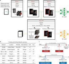
          Millions of breast imaging exams are performed each year in an effort to reduce the morbidity and mortality of breast cancer. Breast imaging exams are performed for cancer screening, diagnostic work-up of suspicious findings, evaluating extent of disease in recently diagnosed breast cancer patients, and determining treatment response. Yet, the interpretation of breast imaging can be subjective, tedious, time-consuming, and prone to human error. Retrospective and small reader studies suggest that deep learning (DL) has great potential to perform medical imaging tasks at or above human-level performance, and may be used to automate aspects of the breast cancer screening process, improve cancer detection rates, decrease unnecessary callbacks and biopsies, optimize patient risk assessment, and open up new possibilities for disease prognostication. Prospective trials are urgently needed to validate these proposed tools, paving the way for real-world clinical use. New regulatory frameworks must also be developed to address the unique ethical, medicolegal, and quality control issues that DL algorithms present. In this article, we review the basics of DL, describe recent DL breast imaging applications including cancer detection and risk prediction, and discuss the challenges and future directions of artificial intelligence-based systems in the field of breast cancer.

          Related collections

          Most cited references96

          • Record: found
          • Abstract: found
          • Article: not found

          Deep learning.

          Deep learning allows computational models that are composed of multiple processing layers to learn representations of data with multiple levels of abstraction. These methods have dramatically improved the state-of-the-art in speech recognition, visual object recognition, object detection and many other domains such as drug discovery and genomics. Deep learning discovers intricate structure in large data sets by using the backpropagation algorithm to indicate how a machine should change its internal parameters that are used to compute the representation in each layer from the representation in the previous layer. Deep convolutional nets have brought about breakthroughs in processing images, video, speech and audio, whereas recurrent nets have shone light on sequential data such as text and speech.
            Bookmark
            • Record: found
            • Abstract: found
            • Article: not found

            Dermatologist-level classification of skin cancer with deep neural networks

            Skin cancer, the most common human malignancy, is primarily diagnosed visually, beginning with an initial clinical screening and followed potentially by dermoscopic analysis, a biopsy and histopathological examination. Automated classification of skin lesions using images is a challenging task owing to the fine-grained variability in the appearance of skin lesions. Deep convolutional neural networks (CNNs) show potential for general and highly variable tasks across many fine-grained object categories. Here we demonstrate classification of skin lesions using a single CNN, trained end-to-end from images directly, using only pixels and disease labels as inputs. We train a CNN using a dataset of 129,450 clinical images—two orders of magnitude larger than previous datasets—consisting of 2,032 different diseases. We test its performance against 21 board-certified dermatologists on biopsy-proven clinical images with two critical binary classification use cases: keratinocyte carcinomas versus benign seborrheic keratoses; and malignant melanomas versus benign nevi. The first case represents the identification of the most common cancers, the second represents the identification of the deadliest skin cancer. The CNN achieves performance on par with all tested experts across both tasks, demonstrating an artificial intelligence capable of classifying skin cancer with a level of competence comparable to dermatologists. Outfitted with deep neural networks, mobile devices can potentially extend the reach of dermatologists outside of the clinic. It is projected that 6.3 billion smartphone subscriptions will exist by the year 2021 (ref. 13) and can therefore potentially provide low-cost universal access to vital diagnostic care.
              Bookmark
              • Record: found
              • Abstract: found
              • Article: not found

              A survey on deep learning in medical image analysis

              Deep learning algorithms, in particular convolutional networks, have rapidly become a methodology of choice for analyzing medical images. This paper reviews the major deep learning concepts pertinent to medical image analysis and summarizes over 300 contributions to the field, most of which appeared in the last year. We survey the use of deep learning for image classification, object detection, segmentation, registration, and other tasks. Concise overviews are provided of studies per application area: neuro, retinal, pulmonary, digital pathology, breast, cardiac, abdominal, musculoskeletal. We end with a summary of the current state-of-the-art, a critical discussion of open challenges and directions for future research.
                Bookmark

                Author and article information

                Contributors
                Journal
                BJR Open
                BJR Open
                bjro
                BJR Open
                The British Institute of Radiology.
                2513-9878
                2022
                13 May 2022
                : 4
                : 1
                : 20210060
                Affiliations
                [1]org-divisionDepartment of Radiology, Memorial Sloan Kettering Cancer Center , New York, NY 10065, United States
                Author notes
                Address correspondence to: Dr Sarah Eskreis-Winkler. E-mail: eskreiss@ 123456mskcc.org
                Article
                BJRO-D-21-00060
                10.1259/bjro.20210060
                9459862
                36105427
                a124ec56-874a-4d65-b9c7-7f72813548da
                © 2022 The Authors. Published by the British Institute of Radiology

                This is an open access article distributed under the terms of the Creative Commons Attribution 4.0 International License, which permits unrestricted use, distribution and reproduction in any medium, provided the original author and source are credited.

                History
                : 05 October 2021
                : 04 April 2022
                : 21 April 2022
                Page count
                Figures: 5, Tables: 0, Equations: 0, References: 95, Pages: 12, Words: 8233
                Categories
                Review Article
                bjro, BJRO
                oth, AI and machine learning
                brst, Breast

                Comments

                Comment on this article