44
views
0
recommends
+1 Recommend
1 collections
    0
    shares

      To submit to Bentham Journals, please click here

      • Record: found
      • Abstract: found
      • Article: found
      Is Open Access

      Development Of A Bio-Inspired Mechatronic Chest Wall Simulator For Evaluating The Performances Of Opto-Electronic Plethysmography

      research-article

      Read this article at

      Bookmark
          There is no author summary for this article yet. Authors can add summaries to their articles on ScienceOpen to make them more accessible to a non-specialist audience.

          Abstract

          Instrumented gait analysis based on optoelectronic systems is an expensive technique used to objectively measure the human movement features and it is generally considered as the gold standard. Opto-electronic plethysmography (OEP) is a particular motion analysis system able to: (i) determine chest wall kinematic via the evaluation of marker displacements placed on the thorax and (ii) compute respiratory volumes during breathing.

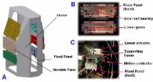
          The aim of this work is to describe the performances of a custom made, bio-inspired, mechatronic chest wall simulator (CWS), specifically designed to assess the metrological performances of the OEP system. The design of the simulator is based on the chest wall kinematic analysis of three healthy subjects previously determined.

          Two sets of experiments were carried out: (i) to investigate the CWS dynamic response using different target displacements (1 - 12 mm), and (ii) to assess the CWS accuracy and precision in simulating quite breathing, covering the physiological range of respiratory frequency and tidal volume.

          Results show that the CWS allows simulating respiratory frequency up to ~ 60 bpm. The difference between the actual displacement and the set one is always < 9 μm. The precision error, expressed as the ratio between measurement uncertainty and the actual displacement, is lower than 0.32 %.

          The observed good performances permit to consider the CWS prototype feasible to be employed for assessing the performances of OEP system in periodical validation routines.

          Related collections

          Most cited references45

          • Record: found
          • Abstract: not found
          • Article: not found

          Measurement of the separate volume changes of rib cage and abdomen during breathing.

            Bookmark
            • Record: found
            • Abstract: found
            • Article: not found

            Chest wall and lung volume estimation by optical reflectance motion analysis.

            Estimation of chest wall motion by surface measurements only allows one-dimensional measurements of the chest wall. We have assessed on optical reflectance system (OR), which tracks reflective markers in three dimensions (3-D) for respiratory use. We used 86 (6-mm-diameter) hemispherical reflective markers arranged circumferentially on the chest wall in seven rows between the sternal notch and the anterior superior iliac crest in two normal standing subjects. We calculated the volume of the entire chest wall and compared inspired and expired volumes with volumes obtained by spirometry. Marker positions were recorded by four TV cameras; two were 4 m in front of and two were 4 m behind the subject. The TV signals were sampled at 100 Hz and combined with grid calibration parameters on a personal computer to obtain the 3-D coordinates of the markers. Chest wall surfaces were reconstructed by triangulation through the point data, and chest wall volume was calculated. During tidal breathing and vital capacity maneuvers and during CO2-stimulated hyperpnea, there was a very close correlation of the lung volumes (VL) estimated by spirometry [VL(SP)] and OR [VL(OR)]. Regression equations of VL(OR) (y) vs. VL(SP) (x, BTPS in liters) for the two subjects were given by y = 1.01x-0.01 (r = 0.996) and y = 0.96x + 0.03 (r = 0.997), and by y = 1.04x + 0.25 (r = 0.97) and y = 0.98x + 0.14 (r = 0.95) for the two maneuvers, respectively. We conclude spirometric volumes can be estimated very accurately and directly from chest wall surface markers, and we speculate that OR may be usefully applied to calculations of chest wall shape, regional volumes, and motion analysis.
              Bookmark
              • Record: found
              • Abstract: found
              • Article: not found

              Regional chest wall volumes during exercise in chronic obstructive pulmonary disease.

              Dynamic hyperinflation of the lungs impairs exercise performance in chronic obstructive pulmonary disease (COPD). However, it is unclear which patients are affected by dynamic hyperinflation and how the respiratory muscles respond to the change in lung volume. Using optoelectronic plethysmography, total and regional chest wall volumes were measured non-invasively in 20 stable patients with COPD (mean (SD) forced expiratory volume in 1 second 43.6 (11.6)% predicted) and dynamic hyperinflation was tracked breath by breath to test if this was the mechanism of exercise limitation. Resting ventilation, breathing pattern, symptoms, rib cage and abdominal volumes were recorded at rest and during symptom limited cycle ergometry. Pleural, abdominal, and transdiaphragmatic pressures were measured in eight patients. End expiratory chest wall volume increased by a mean (SE) of 592 (80) ml in 12 patients (hyperinflators) but decreased by 462 (103) ml in eight (euvolumics). During exercise, tidal volume increased in euvolumic patients by reducing end expiratory abdominal volume while in hyperinflators tidal volume increased by increasing end inspiratory abdominal and rib cage volumes. The maximal abdominal pressure was 22.1 (9.0) cm H(2)O in euvolumic patients and 7.6 (2.6) cm H(2)O in hyperinflators. Euvolumic patients were as breathless as hyperinflators but exercised for less time and reached lower maximum workloads (p<0.05) despite having better spirometric parameters and a greater expiratory flow reserve. Dynamic hyperinflation is not the only mechanism limiting exercise performance in patients with stable COPD. Accurate measurement of chest wall volume can identify the different patterns of respiratory muscle activation during exercise.
                Bookmark

                Author and article information

                Journal
                Open Biomed Eng J
                Open Biomed Eng J
                TOBEJ
                The Open Biomedical Engineering Journal
                Bentham Open
                1874-1207
                19 December 2014
                2014
                : 8
                : 120-130
                Affiliations
                [1 ]Unit of Measurements and Biomedical Instrumentation, Center for Integrated Research, Università Campus Bio-Medico di Roma, Via Álvaro del Portillo, 21, 00128 Rome, Italy
                [2 ]Department of Engineering, University of ROMA TRE, Via della Vasca Navale 79/81, Roma, Italy
                Author notes
                [* ]Address correspondence to the author at the Unit of Measurements and Biomedical Instrumentation, Center for Integrated Research, Università Campus Bio-Medico di Roma, Via Álvaro del Portillo, 21, 00128 Rome, Italy; Tel. +39-06-225419650; E-mail: c.massaroni@ 123456unicampus.it
                Article
                TOBEJ-8-120
                10.2174/1874120701408010120
                4302486
                fa0d99ab-bf2a-4d2a-b698-d375d727b6c4
                © Massaroni et al.; Licensee Bentham Open.

                This is an open access article licensed under the terms of the Creative Commons Attribution Non-Commercial License ( http://creativecommons.org/licenses/by-nc/3.0/) which permits unrestricted, non-commercial use, distribution and reproduction in any medium, provided the work is properly cited.

                History
                : 10 September 2014
                : 29 October 2014
                : 31 October 2014
                Categories
                Article

                Biomedical engineering
                breathing pattern,chest wall kinematic,chest wall simulator,metrological assessment,motion analysis,opto-electronic plethysmography,respiratory monitoring.

                Comments

                Comment on this article