1
views
0
recommends
+1 Recommend
0 collections
    0
    shares
      • Record: found
      • Abstract: found
      • Article: found
      Is Open Access

      Translating microcalcification biomarker information into the laboratory: A preliminary assessment utilizing core biopsies obtained from sites of mammographic calcification

      research-article

      Read this article at

      Bookmark
          There is no author summary for this article yet. Authors can add summaries to their articles on ScienceOpen to make them more accessible to a non-specialist audience.

          Abstract

          Rationale and objectives

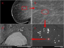
          The potential of breast microcalcification chemistry to provide clinically valuable intelligence is being increasingly studied. However, acquisition of crystallographic details has, to date, been limited to high brightness, synchrotron radiation sources. This study, for the first time, evaluates a laboratory-based system that interrogates histological sections containing microcalcifications. The principal objective was to determine the measurement precision of the laboratory system and assess whether this was sufficient to provide potentially clinical valuable information.

          Materials and methods

          Sections from 5 histological specimens from breast core biopsies obtained to evaluate mammographic calcification were examined using a synchrotron source and a laboratory-based instrument. The samples were chosen to represent a significant proportion of the known breast tissue, mineralogical landscape. Data were subsequently analysed using conventional methods and microcalcification characteristics such as crystallographic phase, chemical deviation from ideal stoichiometry and microstructure were determined.

          Results

          The crystallographic phase of each microcalcification (e.g., hydroxyapatite, whitlockite) was easily determined from the laboratory derived data even when a mixed phase was apparent. Lattice parameter values from the laboratory experiments agreed well with the corresponding synchrotron values and, critically, were determined to precisions that were significantly greater than required for potential clinical exploitation.

          Conclusion

          It has been shown that crystallographic characteristics of microcalcifications can be determined in the laboratory with sufficient precision to have potential clinical value. The work will thus enable exploitation acceleration of these latent microcalcification features as current dependence upon access to limited synchrotron resources is minimized.

          Related collections

          Most cited references16

          • Record: found
          • Abstract: found
          • Article: not found

          Indexing of powder diffraction patterns by iterative use of singular value decomposition

          A. Coelho (2003)
          A fast method for indexing powder diffraction patterns has been developed for large and small lattices of all symmetries. The method is relatively insensitive to impurity peaks and missing highd-spacings: on simulated data, little effect in terms of successful indexing has been observed when one in threed-spacings are randomly removed. Comparison with three of the most popular indexing programs, namelyITO,DICVOL91andTREOR90, has shown that the present method as implemented in the programTOPASis more successful at indexing simulated data. Also significant is that the present method performs well on typically noisy data with large diffractometer zero errors. Critical to its success, the present method uses singular value decomposition in an iterative manner for solving linear equations relatinghklvalues tod-spacings.
            Bookmark
            • Record: found
            • Abstract: found
            • Article: not found
            Is Open Access

            The clinical value of detecting microcalcifications on a mammogram

            Many breast lesions are associated with microcalcifications that are detectable by mammography. In most cases, radiologists are able to distinguish calcifications usually associated with benign diseases from those associated with malignancy. In addition to their value in the early detection of breast carcinoma and accurate radiological diagnosis, the presence of microcalcifications often affects the extent of surgical intervention. Certain types of microcalcifications are associated with negative genetic and molecular characteristics of the tumor and unfavorable prognosis. Microcalcifications localized in the larger ducts (duct-centric, casting-type microcalcifications) represent an independent negative prognostic marker compared to lesions containing other types of microcalcifications and to non-calcified lesions. In this review, we summarize the theoretical and methodological background for understanding the clinical impact and discuss the diagnostic and prognostic value of microcalcifications detected in the breast by mammography.
              Bookmark
              • Record: found
              • Abstract: found
              • Article: not found

              New relationships between breast microcalcifications and cancer

              Background: Breast microcalcifications are key diagnostically significant radiological features for localisation of malignancy. This study explores the hypothesis that breast calcification composition is directly related to the local tissue pathological state. Methods: A total of 236 human breast calcifications from 110 patients were analysed by mid-Fouries transform infrared (FTIR) spectroscopy from three different pathology types (112 invasive carcinoma (IC), 64 in-situ carcinomas and 60 benign). The biochemical composition and the incorporation of carbonate into the hydroxyapatite lattice of the microcalcifications were studied by infrared microspectroscopy. This allowed the spectrally identified composition to be directly correlated with the histopathology grading of the surrounding tissue. Results: The carbonate content of breast microcalcifications was shown to significantly decrease when progressing from benign to malignant disease. In this study, we report significant correlations (P<0.001) between microcalcification chemical composition (carbonate content and protein matrix : mineral ratios) and distinct pathology grades (benign, in-situ carcinoma and ICs). Furthermore, a significant correlation (P<0.001) was observed between carbonate concentrations and carcinoma in-situ sub-grades. Using the two measures of pathology-specific calcification composition (carbonate content and protein matrix : mineral ratios) as the inputs to a two-metric discriminant model sensitivities of 79, 84 and 90% and specificities of 98, 82 and 96% were achieved for benign, ductal carcinoma in situ and invasive malignancies, respectively. Conclusions: We present the first demonstration of a direct link between the chemical nature of microcalcifications and the grade of the pathological breast disease. This suggests that microcalcifications have a significant association with cancer progression, and could be used for future objective analytical classification of breast pathology. A simple two-metric model has been demonstrated, more complex spectral analysis may yeild greater discrimination performance. Furthermore there appears to be a sequential progression of calcification composition.
                Bookmark

                Author and article information

                Contributors
                Journal
                Heliyon
                Heliyon
                Heliyon
                Elsevier
                2405-8440
                11 March 2024
                30 March 2024
                11 March 2024
                : 10
                : 6
                : e27686
                Affiliations
                [a ]Gloucestershire Hospitals NHS Foundation Trust, Cheltenham, United Kingdom
                [b ]Cranfield Forensic Institute, Cranfield University, Swindon, United Kingdom
                [c ]School of Physics and Astronomy, University of Exeter, Exeter, United Kingdom
                [d ]School of Chemical and Physical Sciences, Keele University, Staffordshire, United Kingdom
                [e ]School of Cancer and Pharmaceutical Sciences, King's College London, London, United Kingdom
                Author notes
                [* ]Corresponding author. k.d.rogers@ 123456cranfield.ac.uk
                Article
                S2405-8440(24)03717-4 e27686
                10.1016/j.heliyon.2024.e27686
                10950651
                38509936
                f807c390-4c1e-4784-9382-fa87cc51c52c
                © 2024 Published by Elsevier Ltd.

                This is an open access article under the CC BY-NC-ND license (http://creativecommons.org/licenses/by-nc-nd/4.0/).

                History
                : 3 June 2023
                : 4 March 2024
                : 5 March 2024
                Categories
                Research Article

                microcalcification,x-ray scatter,crystallography,biomarker

                Comments

                Comment on this article