33
views
0
recommends
+1 Recommend
0 collections
    0
    shares
      • Record: found
      • Abstract: found
      • Article: not found

      Integrating Brain Implants With Local and Distributed Computing Devices: A Next Generation Epilepsy Management System

      research-article
      , , , , , , , , , , , , , , , , , , , , , , , , , ,
      IEEE Journal of Translational Engineering in Health and Medicine
      IEEE
      Epilepsy, deep brain stimulation, implantable devices, seizure detection, seizure prediction, distributed computing

      Read this article at

      ScienceOpenPublisherPMC
      Bookmark
          There is no author summary for this article yet. Authors can add summaries to their articles on ScienceOpen to make them more accessible to a non-specialist audience.

          Abstract

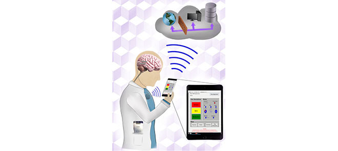
          Brain stimulation has emerged as an effective treatment for a wide range of neurological and psychiatric diseases. Parkinson’s disease, epilepsy, and essential tremor have FDA indications for electrical brain stimulation using intracranially implanted electrodes. Interfacing implantable brain devices with local and cloud computing resources have the potential to improve electrical stimulation efficacy, disease tracking, and management. Epilepsy, in particular, is a neurological disease that might benefit from the integration of brain implants with off-the-body computing for tracking disease and therapy. Recent clinical trials have demonstrated seizure forecasting, seizure detection, and therapeutic electrical stimulation in patients with drug-resistant focal epilepsy. In this paper, we describe a next-generation epilepsy management system that integrates local handheld and cloud-computing resources wirelessly coupled to an implanted device with embedded payloads (sensors, intracranial EEG telemetry, electrical stimulation, classifiers, and control policy implementation). The handheld device and cloud computing resources can provide a seamless interface between patients and physicians, and realtime intracranial EEG can be used to classify brain state (wake/sleep, preseizure, and seizure), implement control policies for electrical stimulation, and track patient health. This system creates a flexible platform in which low demand analytics requiring fast response times are embedded in the implanted device and more complex algorithms are implemented in offthebody local and distributed cloud computing environments. The system enables tracking and management of epileptic neural networks operating over time scales ranging from milliseconds to months.

          Abstract

          Brain Implants integrated with Local and Distributed Computing Devices provide a seamless interface between patients and physicians, and real-time intracranial EEG can be used to classify brain state (wake/sleep, pre-seizure, seizure), implement control policies for electrical stimulation, and track patient health. The system enables tracking and management of epileptic neural networks operating over time scales ranging from milliseconds to months.

          Related collections

          Most cited references41

          • Record: found
          • Abstract: found
          • Article: not found

          Prediction of seizure likelihood with a long-term, implanted seizure advisory system in patients with drug-resistant epilepsy: a first-in-man study.

          Seizure prediction would be clinically useful in patients with epilepsy and could improve safety, increase independence, and allow acute treatment. We did a multicentre clinical feasibility study to assess the safety and efficacy of a long-term implanted seizure advisory system designed to predict seizure likelihood and quantify seizures in adults with drug-resistant focal seizures. We enrolled patients at three centres in Melbourne, Australia, between March 24, 2010, and June 21, 2011. Eligible patients had between two and 12 disabling partial-onset seizures per month, a lateralised epileptogenic zone, and no history of psychogenic seizures. After devices were surgically implanted, patients entered a data collection phase, during which an algorithm for identification of periods of high, moderate, and low seizure likelihood was established. If the algorithm met performance criteria (ie, sensitivity of high-likelihood warnings greater than 65% and performance better than expected through chance prediction of randomly occurring events), patients then entered an advisory phase and received information about seizure likelihood. The primary endpoint was the number of device-related adverse events at 4 months after implantation. Our secondary endpoints were algorithm performance at the end of the data collection phase, clinical effectiveness (measures of anxiety, depression, seizure severity, and quality of life) 4 months after initiation of the advisory phase, and longer-term adverse events. This trial is registered with ClinicalTrials.gov, number NCT01043406. We implanted 15 patients with the advisory system. 11 device-related adverse events were noted within four months of implantation, two of which were serious (device migration, seroma); an additional two serious adverse events occurred during the first year after implantation (device-related infection, device site reaction), but were resolved without further complication. The device met enabling criteria in 11 patients upon completion of the data collection phase, with high likelihood performance estimate sensitivities ranging from 65% to 100%. Three patients' algorithms did not meet performance criteria and one patient required device removal because of an adverse event before sufficient training data were acquired. We detected no significant changes in clinical effectiveness measures between baseline and 4 months after implantation. This study showed that intracranial electroencephalographic monitoring is feasible in ambulatory patients with drug-resistant epilepsy. If these findings are replicated in larger, longer studies, accurate definition of preictal electrical activity might improve understanding of seizure generation and eventually lead to new management strategies. NeuroVista. Copyright © 2013 Elsevier Ltd. All rights reserved.
            Bookmark
            • Record: found
            • Abstract: found
            • Article: not found

            A phase I trial of deep brain stimulation of memory circuits in Alzheimer's disease.

            Alzheimer disease (AD) is characterized by functional impairment in the neural elements and circuits underlying cognitive and memory functions. We hypothesized that fornix/hypothalamus deep brain stimulation (DBS) could modulate neurophysiological activity in these pathological circuits and possibly produce clinical benefits. We conducted a phase I trial in 6 patients with mild AD receiving ongoing medication treatment. Patients received continuous stimulation for 12 months. Three main lines of investigation were pursued including: (1) mapping the brain areas whose physiological function was modulated by stimulation using standardized low-resolution electromagnetic tomography, (2) assessing whether DBS could correct the regional alterations in cerebral glucose metabolism in AD using positron emission tomography (PET), and 3) measuring the effects of DBS on cognitive function over time using clinical scales and instruments. DBS drove neural activity in the memory circuit, including the entorhinal, and hippocampal areas and activated the brain's default mode network. PET scans showed an early and striking reversal of the impaired glucose utilization in the temporal and parietal lobes that was maintained after 12 months of continuous stimulation. Evaluation of the Alzheimer's Disease Assessment Scale cognitive subscale and the Mini Mental State Examination suggested possible improvements and/or slowing in the rate of cognitive decline at 6 and 12 months in some patients. There were no serious adverse events. There is an urgent need for novel therapeutic approaches for AD. Modulating pathological brain activity in this illness with DBS merits further investigation.
              Bookmark
              • Record: found
              • Abstract: found
              • Article: not found

              Long-term seizure outcomes following epilepsy surgery: a systematic review and meta-analysis.

              Assessment of long-term outcomes is essential in brain surgery for epilepsy, which is an irreversible intervention for a chronic condition. Excellent short-term results of resective epilepsy surgery have been established, but less is known about long-term outcomes. We performed a systematic review and meta-analysis of the evidence on this topic. To provide evidence-based estimates of long-term results of various types of epilepsy surgery and to identify sources of variation in results of published studies, we searched Medline, Index Medicus, the Cochrane database, bibliographies of reviews, original articles and book chapters to identify articles published since 1991 that contained > or =20 patients of any age, undergoing resective or non-resective epilepsy surgery, and followed for a mean/median of > or =5 years. Two reviewers independently assessed study eligibility and extracted data, resolving disagreements through discussion. Seventy-six articles fulfilled our eligibility criteria, of which 71 reported on resective surgery (93%) and five (7%) on non-resective surgery. There were no randomized trials and only six studies had a control group. Some articles contributed more than one study, yielding 83 studies of which 78 dealt with resective surgery and five with non-resective surgery. Forty studies (51%) of resective surgery referred to temporal lobe surgery, 25 (32%) to grouped temporal and extratemporal surgery, seven (9%) to frontal surgery, two (3%) to grouped extratemporal surgery, two (3%) to hemispherectomy, and one (1%) each to parietal and occipital surgery. In the non-resective category, three studies reported outcomes after callosotomy and two after multiple subpial transections. The median proportion of long-term seizure-free patients was 66% with temporal lobe resections, 46% with occipital and parietal resections, and 27% with frontal lobe resections. In the long term, only 35% of patients with callosotomy were free of most disabling seizures, and 16% with multiple subpial transections remained free of all seizures. The year of operation, duration of follow-up and outcome classification system were most strongly associated with outcomes. Almost all long-term outcome studies describe patient cohorts without controls. Although there is substantial variation in outcome definition and methodology among the studies, consistent patterns of results emerge for various surgical interventions after adjusting for sources of heterogeneity. The long-term (> or =5 years) seizure free rate following temporal lobe resective surgery was similar to that reported in short-term controlled studies. On the other hand, long-term seizure freedom was consistently lower after extratemporal surgery and palliative procedures.
                Bookmark

                Author and article information

                Contributors
                Journal
                IEEE J Transl Eng Health Med
                IEEE J Transl Eng Health Med
                0063400
                JTEHM
                IJTEBN
                IEEE Journal of Translational Engineering in Health and Medicine
                IEEE
                2168-2372
                2018
                26 September 2018
                : 6
                : 2500112
                Affiliations
                [1]divisionMayo Systems Electrophysiology Laboratory, departmentDepartment of Neurology, institutionMayo Clinic; RochesterMN55905USA
                [2]institutionCzech Institute of Informatics, Robotics and Cybernetics, Czech Technical University in Prague; 160 00PrahaCzech Republic
                [3]departmentDepartment of Physiology and Biomedical Engineering, institutionMayo Clinic; RochesterMN55905USA
                [4]divisionInternational Clinical Research Center, institutionSt. Anne’s University Hospital; 656 91BrnoCzech Republic
                [5]departmentDepartment of Neurosurgery, institutionMayo Clinic; RochesterMN55905USA
                [6]divisionResearch and Core Technology, institutionRestorative Therapy Group, Medtronic; MinneapolisMN55432-3568USA
                [7]divisionCenter for Neuroengineering and Therapeutics, departmentDepartment of Bioengineering, institutionUniversity of Pennsylvania; PhiladelphiaPA19104USA
                [8]departmentDepartment of Veterinary Clinical Sciences, institutionUniversity of Minnesota College of Veterinary Medicine; St. PaulMN55108USA
                [9]departmentDepartment of Surgical and Radiological Sciences, institutionUniversity of California at Davis; DavisCA95616USA
                [10]departmentDepartment of Neurology, institutionMayo Clinic; RochesterMN55905USA
                [11]divisionVeterinary Medical Teaching Hospital, institutionUniversity of California at Davis; DavisCA95616USA
                Author notes
                Article
                2500112
                10.1109/JTEHM.2018.2869398
                6170139
                30310759
                f7ed89b9-89b3-486a-abc1-47869ed09a84
                2168-2372 © 2018 IEEE. Translations and content mining are permitted for academic research only. Personal use is also permitted, but republication/redistribution requires IEEE permission. See http://www.ieee.org/publications_standards/publications/rights/index.html for more information.
                History
                : 02 January 2018
                : 18 June 2018
                : 15 August 2018
                : 16 August 2018
                : 26 September 2018
                Page count
                Figures: 6, Tables: 2, Equations: 4, References: 50, Pages: 12
                Funding
                Funded by: Mayo Clinic, fundref 10.13039/100000871;
                Funded by: NIH Clinical Center, fundref 10.13039/100000098;
                Award ID: R01 NS09288203
                Award ID: UH2/UH3NS95495
                Funded by: České Vysoké Učení Technické v Praze, fundref 10.13039/100007655;
                Funded by: Czech Science Foundation;
                Award ID: 1720480S
                Funded by: Grantová Agentura České Republiky, fundref 10.13039/501100001824;
                Award ID: P103/11/0933
                Funded by: European Regional Development Fund, fundref 10.13039/501100008530;
                Award ID: CZ.1.05/ 1.1.00/02.0123
                Funded by: Mirowski Family Foundation;
                This work was supported in part by the Mayo Clinic Discovery Translation Grant, National Institutes of Health under Grant R01 NS09288203 and Grant UH2/UH3NS95495, the Institutional Resources for Research by Czech Technical University in Prague, Czech Republic, the Czech Science Foundation under Grant 1720480S, the Temporal Context in Analysis of LongTerm NonStationary Multidimensional Signal, Czech Republic Grant Agency, under Grant P103/11/0933, the European Regional Development Fund through the Project FNUSA-ICRC under Grant CZ.1.05/ 1.1.00/02.0123, and the Mirowski Family Foundation.
                Categories
                Article

                epilepsy,deep brain stimulation,implantable devices,seizure detection,seizure prediction,distributed computing

                Comments

                Comment on this article