38
views
0
recommends
+1 Recommend
0 collections
    0
    shares
      • Record: found
      • Abstract: found
      • Article: found
      Is Open Access

      Video capsule endoscopy vs double-balloon enteroscopy in the diagnosis of small bowel bleeding: A systematic review and meta-analysis

      research-article

      Read this article at

      Bookmark
          There is no author summary for this article yet. Authors can add summaries to their articles on ScienceOpen to make them more accessible to a non-specialist audience.

          Abstract

          AIM

          To compare the diagnostic accuracy of video capsule endoscopy (VCE) and double-balloon enteroscopy (DBE) in cases of obscure gastrointestinal bleeding (OGIB) of vascular origin.

          METHODS

          MEDLINE ( via PubMed), LILACS ( via BVS) and Cochrane/CENTRAL virtual databases were searched for studies dated before 2017. We identified prospective and retrospective studies, including observational, cohort, single-blinded and multicenter studies, comparing VCE and DBE for the diagnosis of OGIB, and data of all the vascular sources of bleeding were collected. All patients were subjected to the same gold standard method. Relevant data were then extracted from each included study using a standardized extraction form. We calculated study variables (sensitivity, specificity, prevalence, positive and negative predictive values and accuracy) and performed a meta-analysis using Meta-Disc software.

          RESULTS

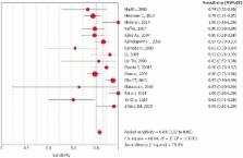
          In the per-patient analysis, 17 studies (1477 lesions) were included. We identified 3150 exams (1722 VCE and 1428 DBE) in 2043 patients and identified 2248 sources of bleeding, 1467 of which were from vascular lesions. Of these lesions, 864 (58.5%) were diagnosed by VCE, and 613 (41.5%) were diagnosed by DBE. The pretest probability for bleeding of vascular origin was 54.34%. The sensitivity of DBE was 84% (95%CI: 0.82-0.86; heterogeneity: 78.00%), and the specificity was 92% (95%CI: 0.89-0.94; heterogeneity: 92.0%). For DBE, the positive likelihood ratio was 11.29 (95%CI: 4.83-26.40; heterogeneity: 91.6%), and the negative likelihood ratio was 0.20 (95%CI: 0.15-0.27; heterogeneity: 67.3%). Performing DBE after CE increased the diagnostic yield of vascular lesion by 7%, from 83% to 90%.

          CONCLUSION

          The diagnostic accuracy of detecting small bowel bleeding from a vascular source is increased with the use of an isolated video capsule endoscope compared with isolated DBE. However, concomitant use increases the detection rate of the bleeding source.

          Related collections

          Most cited references39

          • Record: found
          • Abstract: found
          • Article: not found

          Wireless capsule endoscopy.

          Over the last decade, WCE has established itself as a valuable test for imaging the small intestine. It is a safe and relatively easy procedure to perform that can provide valuable information in the diagnosis of small-bowel conditions. Its applications still remain limited within the esophagus and colon. Future developments may include improving visualization within the esophagus and developing technologies that may allow manipulation of the capsule within the GI tract and biopsy capabilities.
            Bookmark
            • Record: found
            • Abstract: found
            • Article: not found

            Diagnostic value of endoscopic capsule in patients with obscure digestive bleeding: blinded comparison with video push-enteroscopy.

            This study was designed to prospectively compare the diagnostic yield of the M2A endoscopic capsule with that of video push-enteroscopy in exploring the small intestine in patients with obscure digestive bleeding. Patients with either occult or overt obscure digestive bleeding and a negative endoscopic work-up underwent a double intestinal investigation, with video push-enteroscopy and a wireless capsule, performed blindly by separate examiners. The diagnostic yield for each technique was defined as the frequency of detection of clinically relevant intestinal lesions, i. e. those having the potential for bleeding. 60 patients (27 men, 33 women; age 58 +/- 18 years; hemoglobin 9.4 +/- 2.5 g/dl) were included, 32 with occult and 28 with overt bleeding. Results were analyzed for 58 patients, who completed both examinations. Lesions were detected in 43 patients: with both techniques in 19, only by capsule in 21, and, conversely, only by push-enteroscopy in 3 ( P = 0.04). Final diagnoses were as follows: a P2 lesion with high bleeding potential in 28 patients (19 angiomata, 6 ulcerations, 2 tumors, 1 case of intestinal varices); a P1 lesion with intermediate bleeding potential in 15 patients (2 patients with mucosal erosions, 13 patients with mucosal red spots); and there were normal findings from 15 procedures. No procedure induced any complication. The use of the wireless endoscopy capsule detects significantly more clinically relevant intestinal lesions than video push-enteroscopy in patients with obscure digestive bleeding, raising the diagnostic yield to 67.2 %. Its influence on the clinical outcome for patients needs further investigation.
              Bookmark
              • Record: found
              • Abstract: found
              • Article: not found

              The role of endoscopy in the management of obscure GI bleeding.

              This is one of a series of statements discussing the use of GI endoscopy in common clinical situations. The Standards of Practice Committee of the American Society for Gastrointestinal Endoscopy (ASGE) prepared this text. In preparing this guideline, we performed a search of the medical literature by using PubMed. Additional references were obtained from the bibliographies of the identified articles and from recommendations of expert consultants. Guidelines for appropriate use of endoscopy are based on a critical review of the available data and expert consensus at the time the guidelines were drafted. Further controlled clinical studies may be needed to clarify aspects of this guideline. This guideline may be revised as necessary to account for changes in technology, new data, or other aspects of clinical practice. The recommendations are based on reviewed studies and are graded on the strength of the supporting evidence (Table 1).(1) The strength of individual recommendations is based both upon the aggregate evidence quality and an assessment of the anticipated benefits and harms. Weaker recommendations are indicated by phrases such as "we suggest," whereas stronger recommendations are typically stated as "we recommend." This guideline is intended to be an educational device to provide information that may assist endoscopists in providing care to patients. This guideline is not a rule and should not be construed as establishing a legal standard of care or as encouraging, advocating, requiring, or discouraging any particular treatment. Clinical decisions in any particular case involve a complex analysis of the patient's condition and available courses of action. Therefore, clinical considerations may lead an endoscopist to take a course of action that varies from these guidelines. Copyright 2010 American Society for Gastrointestinal Endoscopy. Published by Mosby, Inc. All rights reserved.
                Bookmark

                Author and article information

                Contributors
                Journal
                World J Gastrointest Endosc
                WJGE
                World Journal of Gastrointestinal Endoscopy
                Baishideng Publishing Group Inc
                1948-5190
                16 December 2018
                16 December 2018
                : 10
                : 12
                : 400-421
                Affiliations
                Department of Endoscopy of Clinics Hospital of São Paulo University, São Paulo 05403-00, Brazil
                Department of Endoscopy of Clinics Hospital of São Paulo University, São Paulo 05403-00, Brazil. igorbraga1@ 123456gmail.com
                Department of Endoscopy of Clinics Hospital of São Paulo University, São Paulo 05403-00, Brazil
                Department of Endoscopy of Clinics Hospital of São Paulo University, São Paulo 05403-00, Brazil
                Department of Endoscopy of Clinics Hospital of São Paulo University, São Paulo 05403-00, Brazil
                Department of Endoscopy of Clinics Hospital of São Paulo University, São Paulo 05403-00, Brazil
                Molecular and Cell Biology, University of California, California, Berkeley, CA 94720, United States
                Department of Endoscopy of Clinics Hospital of São Paulo University, São Paulo 05403-00, Brazil
                Department of Endoscopy of Clinics Hospital of São Paulo University, São Paulo 05403-00, Brazil
                Department of Endoscopy of Clinics Hospital of São Paulo University, São Paulo 05403-00, Brazil
                Author notes

                Author contributions: Brito HP acquisition of data, analysis, interpretation of data, drafting of the article, revision of the article, final approval; Bernardo WM analysis and interpretation of data, drafting of the article, final approval; Chaves DM acquisition of data, drafting of the article, revision of the article, final approval; de Moura DTH and Ribeiro IB: analysis and interpretation of data, revision of the article; Kuga R acquisition of data, drafting of the article, revision of the article, Maahs ED revision of the article; de Moura ETH drafting of the article, revision of the article; de Moura EGH analysis and interpretation of data, drafting of the article, revision of the article, final approval; de Moura EGH: conception and design of the study, critical revision, final approval.

                Corresponding author to: Igor Braga Ribeiro, MD, Academic Fellow, Surgeon, Gastrointestinal Endoscopy Unit, Hospital das Clínicas da Faculdade de Medicina da Universidade de São Paulo, Av. Dr. Enéas de Carvalho Aguiar 255, Instituto Central, Prédio dos Ambulatórios, Pinheiros, São Paulo 05403-000, Brazil. igorbraga1@ 123456gmail.com

                Telephone: +55-92-981377788

                Article
                jWJGE.v10.i12.pg400
                10.4253/wjge.v10.i12.400
                6323498
                30631404
                f6b98e43-c18f-485b-92a4-8430fb10ce3d
                ©The Author(s) 2018. Published by Baishideng Publishing Group Inc. All rights reserved.

                This article is an open-access article which was selected by an in-house editor and fully peer-reviewed by external reviewers. It is distributed in accordance with the Creative Commons Attribution Non Commercial (CC BY-NC 4.0) license, which permits others to distribute, remix, adapt, build upon this work non-commercially, and license their derivative works on different terms, provided the original work is properly cited and the use is non-commercial.

                History
                : 25 July 2018
                : 31 August 2018
                : 15 November 2018
                Categories
                Meta-Analysis

                small bowel bleeding,hemorrhage,upper gastrointestinal bleeding,obscure hemorrhage,enteroscopy

                Comments

                Comment on this article