6
views
0
recommends
+1 Recommend
0 collections
    0
    shares
      • Record: found
      • Abstract: found
      • Article: found
      Is Open Access

      Topography and distribution of adenosine A 2A and dopamine D 2 receptors in the human Subthalamic Nucleus

      brief-report

      Read this article at

      Bookmark
          There is no author summary for this article yet. Authors can add summaries to their articles on ScienceOpen to make them more accessible to a non-specialist audience.

          Abstract

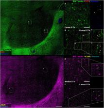
          The human Subthalamic Nucleus (STh) is a diencephalic lens-shaped structure located ventrally to the thalamus and functionally implicated in the basal ganglia circuits. Despite recent efforts to characterize the neurochemical and functional anatomy of the STh, little to no information is available concerning the expression and distribution of receptors belonging to the dopaminergic and purinergic system in the human STh. Both systems are consistently implicated in basal ganglia physiology and pathology, especially in Parkinson’s Disease, and represent important targets for the pharmacological treatment of movement disorders. Here, we investigate the topography and distribution of A 2A adenosine and D 2 dopamine receptors in the human basal ganglia and subthalamic nucleus. Our findings indicate a peculiar topographical distribution of the two receptors throughout the subthalamic nucleus, while colocalization between the receptors opens the possibility for the presence of A 2AR- D 2R heterodimers within the dorsal and medial aspects of the structure. However, further investigation is required to confirm these findings.

          Related collections

          Most cited references43

          • Record: found
          • Abstract: found
          • Article: not found

          Adenosine-dopamine receptor-receptor interactions as an integrative mechanism in the basal ganglia.

          Increasing evidence suggests that antagonistic interactions between specific subtypes of adenosine and dopamine receptors in the basal ganglia are involved in the motor depressant effects of adenosine receptor agonists and the motor stimulant effects of adenosine receptor antagonists, such as caffeine. The GABAergic striatopallidal neurons are regulated by interacting adenosine A2A and dopamine D2 receptors. On the other hand, the GABAergic striatonigral and striatoentopeduncular neurons seem to be regulated by interacting adenosine A1 and dopamine D1 receptors. Furthermore, behavioural studies have revealed interactions between adenosine A2A and dopamine D1 receptors that occur at the network level. These adenosine-dopamine receptor-receptor interactions might offer new therapeutic leads for basal ganglia disorders.
            Bookmark
            • Record: found
            • Abstract: found
            • Article: not found

            Adenosine A2A receptors and basal ganglia physiology.

            Adenosine A2A receptors are highly enriched in the basal ganglia system. They are predominantly expressed in enkephalin-expressing GABAergic striatopallidal neurons and therefore are highly relevant to the function of the indirect efferent pathway of the basal ganglia system. In these GABAergic enkephalinergic neurons, the A2A receptor tightly interacts structurally and functionally with the dopamine D2 receptor. Both by forming receptor heteromers and by targeting common intracellular signaling cascades, A2A and D2 receptors exhibit reciprocal antagonistic interactions that are central to the function of the indirect pathway and hence to basal ganglia control of movement, motor learning, motivation and reward. Consequently, this A2A/D2 receptors antagonistic interaction is also central to basal ganglia dysfunction in Parkinson's disease. However, recent evidence demonstrates that, in addition to this post-synaptic site of action, striatal A2A receptors are also expressed and have physiological relevance on pre-synaptic glutamatergic terminals of the cortico-limbic-striatal and thalamo-striatal pathways, where they form heteromeric receptor complexes with adenosine A1 receptors. Therefore, A2A receptors play an important fine-tuning role, boosting the efficiency of glutamatergic information flow in the indirect pathway by exerting control, either pre- and/or post-synaptically, over other key modulators of glutamatergic synapses, including D2 receptors, group I metabotropic mGlu5 glutamate receptors and cannabinoid CB1 receptors, and by triggering the cAMP-protein kinase A signaling cascade.
              Bookmark
              • Record: found
              • Abstract: found
              • Article: not found

              Targeting adenosine A2A receptors in Parkinson's disease.

              The adenosine A2A receptor has emerged as an attractive non-dopaminergic target in the pursuit of improved therapy for Parkinson's disease (PD), based in part on its unique CNS distribution. It is highly enriched in striatopallidal neurons and can form functional heteromeric complexes with other G-protein-coupled receptors, including dopamine D2, metabotropic glutamate mGlu5 and adenosine A1 receptors. Blockade of the adenosine A2A receptor in striatopallidal neurons reduces postsynaptic effects of dopamine depletion, and in turn lessens the motor deficits of PD. A2A antagonists might partially improve not only the symptoms of PD but also its course, by slowing the underlying neurodegeneration and reducing the maladaptive neuroplasticity that complicates standard 'dopamine replacement' treatments. Thus, we review here a prime example of translational neuroscience, through which antagonism of A2A receptors has now entered the arena of clinical trials with realistic prospects for advancing PD therapeutics.
                Bookmark

                Author and article information

                Contributors
                Journal
                Front Neurosci
                Front Neurosci
                Front. Neurosci.
                Frontiers in Neuroscience
                Frontiers Media S.A.
                1662-4548
                1662-453X
                09 August 2022
                2022
                : 16
                : 945574
                Affiliations
                [1] 1Department of Neurosciences, Institute of Human Anatomy, University of Padova , Padua, Italy
                [2] 2Center for Neurodegenerative Disease Research (CESNE), University of Padova , Padua, Italy
                [3] 3Movement Disorders Unit, Neurology Clinic, University Hospital of Padova , Padua, Italy
                Author notes

                Edited by: Giuseppe Anastasi, University of Messina, Italy

                Reviewed by: Francesco Fornai, University of Pisa, Italy; Manuel Narvaez Peláez, University of Malaga, Spain

                *Correspondence: Andrea Porzionato, andrea.porzionato@ 123456unipd.it

                These authors have contributed equally to this work and share first authorship

                This article was submitted to Neurodegeneration, a section of the journal Frontiers in Neuroscience

                Article
                10.3389/fnins.2022.945574
                9396224
                36017181
                f4727ddc-8716-4ef6-9560-817dce8f05ad
                Copyright © 2022 Emmi, Antonini, Sandre, Baldo, Contran, Macchi, Guidolin, Porzionato and De Caro.

                This is an open-access article distributed under the terms of the Creative Commons Attribution License (CC BY). The use, distribution or reproduction in other forums is permitted, provided the original author(s) and the copyright owner(s) are credited and that the original publication in this journal is cited, in accordance with accepted academic practice. No use, distribution or reproduction is permitted which does not comply with these terms.

                History
                : 16 May 2022
                : 20 July 2022
                Page count
                Figures: 4, Tables: 1, Equations: 0, References: 43, Pages: 10, Words: 5584
                Categories
                Neuroscience
                Brief Research Report

                Neurosciences
                d2r,a2ar,subthalamic nucleus,receptor-receptor interactions,neuroanatomy,parkinson’s disease,deep brain stimulation,neurodegeneration

                Comments

                Comment on this article