29
views
0
recommends
+1 Recommend
0 collections
    0
    shares
      • Record: found
      • Abstract: found
      • Article: not found

      Pitx2 promotes heart repair by activating the antioxidant response after cardiac injury

      research-article

      Read this article at

      ScienceOpenPublisherPMC
      Bookmark
          There is no author summary for this article yet. Authors can add summaries to their articles on ScienceOpen to make them more accessible to a non-specialist audience.

          Summary

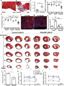
          Myocardial infarction results in compromised myocardial function with heart failure due to insufficient cardiomyocyte self-renewal 1 . Unlike lower vertebrates, mammalian hearts only have a transient neonatal renewal capacity 2 . Reactivating primitive reparative ability in the mature heart requires knowledge of the mechanisms promoting early heart repair. By testing an established Hippo-deficient heart regeneration model for renewal promoting factors, we found that Pitx2 expression was induced in injured, Hippo-deficient ventricles. Pitx2-deficient neonatal hearts failed to repair after apex resection while Pitx2-gain-of-function in adult cardiomyocytes conferred reparative ability after myocardial infarction. Genomic analyses indicated that Pitx2 activated genes encoding electron transport chain components and reactive oxygen species scavengers. A subset of Pitx2 target genes was cooperatively regulated with the Hippo effector, Yap. Furthermore, Nrf2, a regulator of antioxidant response 3 , directly regulated Pitx2 expression and subcellular localization. Pitx2 mutant myocardium had elevated reactive oxygen species levels while antioxidant supplementation suppressed the Pitx2-loss-of-function phenotype. These findings reveal a genetic pathway, activated by tissue damage that is essential for cardiac repair.

          Related collections

          Most cited references13

          • Record: found
          • Abstract: found
          • Article: not found

          The oxygen-rich postnatal environment induces cardiomyocyte cell-cycle arrest through DNA damage response.

          The mammalian heart has a remarkable regenerative capacity for a short period of time after birth, after which the majority of cardiomyocytes permanently exit cell cycle. We sought to determine the primary postnatal event that results in cardiomyocyte cell-cycle arrest. We hypothesized that transition to the oxygen-rich postnatal environment is the upstream signal that results in cell-cycle arrest of cardiomyocytes. Here, we show that reactive oxygen species (ROS), oxidative DNA damage, and DNA damage response (DDR) markers significantly increase in the heart during the first postnatal week. Intriguingly, postnatal hypoxemia, ROS scavenging, or inhibition of DDR all prolong the postnatal proliferative window of cardiomyocytes, whereas hyperoxemia and ROS generators shorten it. These findings uncover a protective mechanism that mediates cardiomyocyte cell-cycle arrest in exchange for utilization of oxygen-dependent aerobic metabolism. Reduction of mitochondrial-dependent oxidative stress should be an important component of cardiomyocyte proliferation-based therapeutic approaches. Copyright © 2014 Elsevier Inc. All rights reserved.
            Bookmark
            • Record: found
            • Abstract: found
            • Article: not found

            Hippo signaling impedes adult heart regeneration.

            Heart failure due to cardiomyocyte loss after ischemic heart disease is the leading cause of death in the United States in large part because heart muscle regenerates poorly. The endogenous mechanisms preventing mammalian cardiomyocyte regeneration are poorly understood. Hippo signaling, an ancient organ size control pathway, is a kinase cascade that inhibits developing cardiomyocyte proliferation but it has not been studied postnatally or in fully mature adult cardiomyocytes. Here, we investigated Hippo signaling in adult cardiomyocyte renewal and regeneration. We found that unstressed Hippo-deficient adult mouse cardiomyocytes re-enter the cell cycle and undergo cytokinesis. Moreover, Hippo deficiency enhances cardiomyocyte regeneration with functional recovery after adult myocardial infarction as well as after postnatal day eight (P8) cardiac apex resection and P8 myocardial infarction. In damaged hearts, Hippo mutant cardiomyocytes also have elevated proliferation. Our findings reveal that Hippo signaling is an endogenous repressor of adult cardiomyocyte renewal and regeneration. Targeting the Hippo pathway in human disease might be beneficial for the treatment of heart disease.
              Bookmark
              • Record: found
              • Abstract: found
              • Article: found
              Is Open Access

              YAP1 Exerts Its Transcriptional Control via TEAD-Mediated Activation of Enhancers

              YAP1 is a major effector of the Hippo pathway and a well-established oncogene. Elevated YAP1 activity due to mutations in Hippo pathway components or YAP1 amplification is observed in several types of human cancers. Here we investigated its genomic binding landscape in YAP1-activated cancer cells, as well as in non-transformed cells. We demonstrate that TEAD transcription factors mediate YAP1 chromatin-binding genome-wide, further explaining their dominant role as primary mediators of YAP1-transcriptional activity. Moreover, we show that YAP1 largely exerts its transcriptional control via distal enhancers that are marked by H3K27 acetylation and that YAP1 is necessary for this chromatin mark at bound enhancers and the activity of the associated genes. This work establishes YAP1-mediated transcriptional regulation at distal enhancers and provides an expanded set of target genes resulting in a fundamental source to study YAP1 function in a normal and cancer setting.
                Bookmark

                Author and article information

                Journal
                0410462
                6011
                Nature
                Nature
                Nature
                0028-0836
                1476-4687
                12 May 2016
                25 May 2016
                2 June 2016
                02 December 2016
                : 534
                : 7605
                : 119-123
                Affiliations
                [1 ]Department of Molecular Physiology and Biophysics, Baylor College of Medicine, Houston, TX 77030, USA
                [2 ]Texas Heart Institute, Houston, TX 77030, USA
                [3 ]Department of Anatomy and Cell Biology and the Craniofacial Anomalies Research Center, The University of Iowa, Iowa City, IA 52242, USA
                [4 ]Department of Molecular Biology and Hamon Center for Regenerative Science and Medicine, University of Texas Southwestern Medical Center, Dallas, TX 75390-9148
                [5 ]Program in Developmental Biology, Baylor College of Medicine, Houston, TX 77030
                [6 ]Cardiovascular Research Institute, Baylor College of Medicine, Houston, TX 77030
                Author notes
                [* ]To whom correspondence should be addressed. jfmartin@ 123456bcm.edu
                Article
                NIHMS773746
                10.1038/nature17959
                4999251
                27251288
                ee4b0d71-6e31-49a4-b2e1-be4ae5b8c46d

                Users may view, print, copy, and download text and data-mine the content in such documents, for the purposes of academic research, subject always to the full Conditions of use: http://www.nature.com/authors/editorial_policies/license.html#terms

                History
                Categories
                Article

                Uncategorized
                Uncategorized

                Comments

                Comment on this article