20
views
0
recommends
+1 Recommend
0 collections
    0
    shares
      • Record: found
      • Abstract: found
      • Article: not found

      Assembly of the Dystrophin-Associated Protein Complex Does Not Require the Dystrophin Cooh-Terminal Domain

      research-article

      Read this article at

      ScienceOpenPublisherPMC
      Bookmark
          There is no author summary for this article yet. Authors can add summaries to their articles on ScienceOpen to make them more accessible to a non-specialist audience.

          Abstract

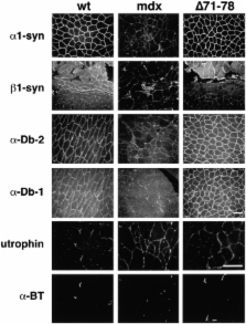
          Dystrophin is a multidomain protein that links the actin cytoskeleton to laminin in the extracellular matrix through the dystrophin associated protein (DAP) complex. The COOH-terminal domain of dystrophin binds to two components of the DAP complex, syntrophin and dystrobrevin. To understand the role of syntrophin and dystrobrevin, we previously generated a series of transgenic mouse lines expressing dystrophins with deletions throughout the COOH-terminal domain. Each of these mice had normal muscle function and displayed normal localization of syntrophin and dystrobrevin. Since syntrophin and dystrobrevin bind to each other as well as to dystrophin, we have now generated a transgenic mouse deleted for the entire dystrophin COOH-terminal domain. Unexpectedly, this truncated dystrophin supported normal muscle function and assembly of the DAP complex. These results demonstrate that syntrophin and dystrobrevin functionally associate with the DAP complex in the absence of a direct link to dystrophin. We also observed that the DAP complexes in these different transgenic mouse strains were not identical. Instead, the DAP complexes contained varying ratios of syntrophin and dystrobrevin isoforms. These results suggest that alternative splicing of the dystrophin gene, which naturally generates COOH-terminal deletions in dystrophin, may function to regulate the isoform composition of the DAP complex.

          Related collections

          Most cited references88

          • Record: found
          • Abstract: found
          • Article: not found

          Dystrophin protects the sarcolemma from stresses developed during muscle contraction.

          The protein dystrophin, normally found on the cytoplasmic surface of skeletal muscle cell membranes, is absent in patients with Duchenne muscular dystrophy as well as mdx (X-linked muscular dystrophy) mice. Although its primary structure has been determined, the precise functional role of dystrophin remains the subject of speculation. In the present study, we demonstrate that dystrophin-deficient muscle fibers of the mdx mouse exhibit an increased susceptibility to contraction-induced sarcolemmal rupture. The level of sarcolemmal damage is directly correlated with the magnitude of mechanical stress placed upon the membrane during contraction rather than the number of activations of the muscle. These findings strongly support the proposition that the primary function of dystrophin is to provide mechanical reinforcement to the sarcolemma and thereby protect it from the membrane stresses developed during muscle contraction. Furthermore, the methodology used in this study should prove useful in assessing the efficacy of dystrophin gene therapy in the mdx mouse.
            Bookmark
            • Record: found
            • Abstract: found
            • Article: not found

            Primary structure of dystrophin-associated glycoproteins linking dystrophin to the extracellular matrix.

            The primary sequence of two components of the dystrophin-glycoprotein complex has been established by complementary, DNA cloning. The transmembrane 43K and extracellular 156K dystrophin-associated glycoproteins (DAGs) are encoded by a single messenger RNA and the extracellular 156K DAG binds laminin. Thus, the 156K DAG is a new laminin-binding glycoprotein which may provide a linkage between the sarcolemma and extracellular matrix. These results support the hypothesis that the dramatic reduction in the 156K DAG in Duchenne muscular dystrophy leads to a loss of a linkage between the sarcolemma and extracellular matrix and that this may render muscle fibres more susceptible to necrosis.
              Bookmark
              • Record: found
              • Abstract: found
              • Article: not found

              The mdx mouse diaphragm reproduces the degenerative changes of Duchenne muscular dystrophy.

              Although murine X-linked muscular dystrophy (mdx) and Duchenne muscular dystrophy (DMD) are genetically homologous and both characterized by a complete absence of dystrophin, the limb muscles of adult mdx mice suffer neither the detectable weakness nor the progressive degeneration that are features of DMD. Here we show that the mdx mouse diaphragm exhibits a pattern of degeneration, fibrosis and severe functional deficit comparable to that of DMD limb muscle, although adult mice show no overt respiratory impairment. Progressive functional changes include reductions in strength (to 13.5% of control by two years of age), elasticity, twitch speed and fibre length. The collagen density rises to at least seven times that of control diaphragm and ten times that of mdx hind-limb muscle. By 1.5 years of age, similar but less severe histological changes emerge in the accessory muscles of respiration. On the basis of these findings, we propose that dystrophin deficiency alters the threshold for work-induced injury. Our data provide a quantitative framework for studying the pathogenesis of dystrophy and extend the application of the mdx mouse as an animal model.
                Bookmark

                Author and article information

                Contributors
                Journal
                J Cell Biol
                The Journal of Cell Biology
                The Rockefeller University Press
                0021-9525
                1540-8140
                18 September 2000
                : 150
                : 6
                : 1399-1410
                Affiliations
                [a ]Program in Cellular and Molecular Biology, University of Michigan, Ann Arbor, Michigan 48109-0618
                [b ]Department of Human Genetics, University of Michigan, Ann Arbor, Michigan 48109-0618
                [c ]Center for Gene Therapy, University of Michigan, Ann Arbor, Michigan 48109-0618
                [d ]Department of Physiology, University of Michigan, Ann Arbor, Michigan 48109-0618
                [e ]Howard Hughes Medical Institute, Department of Physiology and Biophysics, Department of Neurology, University of Iowa College of Medicine, Iowa City, Iowa 52242
                [f ]Department of Cell and Molecular Physiology, University of North Carolina at Chapel Hill, Chapel Hill, North Carolina 27599-7545
                Article
                0005104
                10.1083/jcb.150.6.1399
                2150715
                10995444
                ee164493-512a-4346-8269-136405b492bc
                © 2000 The Rockefeller University Press
                History
                : 18 May 2000
                : 26 July 2000
                : 2 August 2000
                Categories
                Original Article

                Cell biology
                mdx mice,syntrophin,muscular dystrophy,dystrophin,dystrobrevin
                Cell biology
                mdx mice, syntrophin, muscular dystrophy, dystrophin, dystrobrevin

                Comments

                Comment on this article