37
views
0
recommends
+1 Recommend
0 collections
    0
    shares
      • Record: found
      • Abstract: found
      • Article: found
      Is Open Access

      Endothelial Galectin-1 Binds to Specific Glycans on Nipah Virus Fusion Protein and Inhibits Maturation, Mobility, and Function to Block Syncytia Formation

      research-article

      Read this article at

      Bookmark
          There is no author summary for this article yet. Authors can add summaries to their articles on ScienceOpen to make them more accessible to a non-specialist audience.

          Abstract

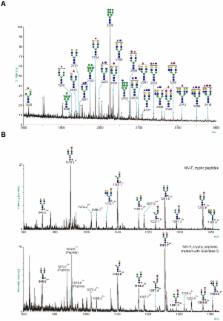
          Nipah virus targets human endothelial cells via NiV-F and NiV-G envelope glycoproteins, resulting in endothelial syncytia formation and vascular compromise. Endothelial cells respond to viral infection by releasing innate immune effectors, including galectins, which are secreted proteins that bind to specific glycan ligands on cell surface glycoproteins. We demonstrate that galectin-1 reduces NiV-F mediated fusion of endothelial cells, and that endogenous galectin-1 in endothelial cells is sufficient to inhibit syncytia formation. Galectin-1 regulates NiV-F mediated cell fusion at three distinct points, including retarding maturation of nascent NiV-F, reducing NiV-F lateral mobility on the plasma membrane, and directly inhibiting the conformational change in NiV-F required for triggering fusion. Characterization of the NiV-F N-glycome showed that the critical site for galectin-1 inhibition is rich in glycan structures known to bind galectin-1. These studies identify a unique set of mechanisms for regulating pathophysiology of NiV infection at the level of the target cell.

          Author Summary

          Nipah virus (NiV) is classified as a “priority pathogen” by the NIH. NiV infection of humans results in multi-organ hemorrhage due to endothelial syncytia formation, and also causes fatal encephalitis in up to 70% of patients. As there are no effective vaccines or therapeutics for NiV, understanding the mechanism of endothelial damage by NiV is a critical goal. Our present work defines the interaction between galectin-1, an innate immune lectin that is secreted by human endothelial cells, with the fusion glycoprotein of NiV. We demonstrate that galectin-1 can block the function of the NiV-F protein via three distinct mechanisms, and thus reduce the ability of NiV-F to cause endothelial cell-cell fusion. Importantly, in this study, we use human endothelial cells, the primary target of Nipah virus in vivo, and demonstrate that endogenous galectin-1 made by endothelial cells contributes to limiting cell-cell fusion caused by NiV-F. As endothelial syncytia formation is one of the primary pathophysiologic events in Nipah virus infection, contributing to the hemorrhagic diathesis seen in infected patients, understanding the mechanism of endothelial cell fusion and the ability of galectin-1 to ameliorate cell fusion are critical for development of new approaches to mitigate these events.

          Related collections

          Most cited references56

          • Record: found
          • Abstract: found
          • Article: not found

          Complex N-glycan number and degree of branching cooperate to regulate cell proliferation and differentiation.

          The number of N-glycans (n) is a distinct feature of each glycoprotein sequence and cooperates with the physical properties of the Golgi N-glycan-branching pathway to regulate surface glycoprotein levels. The Golgi pathway is ultrasensitive to hexosamine flux for the production of tri- and tetra-antennary N-glycans, which bind to galectins and form a molecular lattice that opposes glycoprotein endocytosis. Glycoproteins with few N-glycans (e.g., TbetaR, CTLA-4, and GLUT4) exhibit enhanced cell-surface expression with switch-like responses to increasing hexosamine concentration, whereas glycoproteins with high numbers of N-glycans (e.g., EGFR, IGFR, FGFR, and PDGFR) exhibit hyperbolic responses. Computational and experimental data reveal that these features allow nutrient flux stimulated by growth-promoting high-n receptors to drive arrest/differentiation programs by increasing surface levels of low-n glycoproteins. We have identified a mechanism for metabolic regulation of cellular transition between growth and arrest in mammals arising from apparent coevolution of N-glycan number and branching.
            Bookmark
            • Record: found
            • Abstract: found
            • Article: not found

            EphrinB2 is the entry receptor for Nipah virus, an emergent deadly paramyxovirus.

            Nipah virus (NiV) is an emergent paramyxovirus that causes fatal encephalitis in up to 70 percent of infected patients, and there is evidence of human-to-human transmission. Endothelial syncytia, comprised of multinucleated giant-endothelial cells, are frequently found in NiV infections, and are mediated by the fusion (F) and attachment (G) envelope glycoproteins. Identification of the receptor for this virus will shed light on the pathobiology of NiV infection, and spur the rational development of effective therapeutics. Here we report that ephrinB2, the membrane-bound ligand for the EphB class of receptor tyrosine kinases (RTKs), specifically binds to the attachment (G) glycoprotein of NiV. Soluble Fc-fusion proteins of ephrinB2, but not ephrinB1, effectively block NiV fusion and entry into permissive cell types. Moreover, transfection of ephrinB2 into non-permissive cells renders them permissive for NiV fusion and entry. EphrinB2 is expressed on endothelial cells and neurons, which is consistent with the known cellular tropism for NiV. Significantly, we find that NiV-envelope-mediated infection of microvascular endothelial cells and primary cortical rat neurons is inhibited by soluble ephrinB2, but not by the related ephrinB1 protein. Cumulatively, our data show that ephrinB2 is a functional receptor for NiV.
              Bookmark
              • Record: found
              • Abstract: found
              • Article: not found

              Ephrin-B2 ligand is a functional receptor for Hendra virus and Nipah virus.

              Hendra virus (HeV) and Nipah virus (NiV) belong to the genus Henipavirus of the family Paramyxoviridae and are unique in that they exhibit a broad species tropism and cause fatal disease in both animals and humans. They infect cells through a pH-independent membrane fusion process mediated by their fusion and attachment glycoproteins. Previously, we demonstrated identical cell fusion tropisms for HeV and NiV and the protease-sensitive nature of their unknown cell receptor and identified a human cell line (HeLa-USU) that was nonpermissive for fusion and virus infection. Here, a microarray analysis was performed on the HeLa-USU cells, permissive HeLa-CCL2 cells, and two other permissive human cell lines. From this analysis, we identified a list of genes encoding known and predicted plasma membrane surface-expressed proteins that were highly expressed in all permissive cells and absent from the HeLa-USU cells and rank-ordered them based on their relative levels. Available expression vectors containing the first 10 genes were obtained and individually transfected into HeLa-USU cells. One clone, encoding human ephrin-B2 (EFNB2), was found capable of rendering HeLa-USU cells permissive for HeV- and NiV-mediated cell fusion as well as infection by live virus. A soluble recombinant EFNB2 could potently block fusion and infection and bind soluble recombinant HeV and NiV attachment glycoproteins with high affinity. Together, these data indicate that EFNB2 serves as a functional receptor for both HeV and NiV. The highly conserved nature of EFNB2 in humans and animals is consistent with the broad tropism exhibited by these emerging zoonotic viruses.
                Bookmark

                Author and article information

                Contributors
                Role: Editor
                Journal
                PLoS Pathog
                plos
                plospath
                PLoS Pathogens
                Public Library of Science (San Francisco, USA )
                1553-7366
                1553-7374
                July 2010
                July 2010
                15 July 2010
                : 6
                : 7
                : e1000993
                Affiliations
                [1 ]Department of Pathology and Laboratory Medicine, David Geffen School of Medicine at UCLA, Los Angeles, California, United States of America
                [2 ]Department of Microbiology, Immunology, and Molecular Genetics, David Geffen School of Medicine at UCLA, Los Angeles, California, United States of America
                [3 ]Division of Molecular Biosciences, Faculty of Natural Sciences, Imperial College, London, United Kingdom
                [4 ]MSCAN Ltd., Millars Business Centre, Wokingham, Berks, United Kingdom
                University of California Irvine, United States of America
                Author notes

                Conceived and designed the experiments: OBG HCA JAF ELL SMH HRM AD BL LGB. Performed the experiments: OBG HCA JAF ELL RH LW LRR VA MP. Analyzed the data: OBG HCA JAF ELL RH LW LRR VA MP SMH HRM AD BL LGB. Contributed reagents/materials/analysis tools: OBG HCA JAF SMH HRM. Wrote the paper: OBG HCA SMH HRM AD BL LGB.

                Article
                10-PLPA-RA-2850R2
                10.1371/journal.ppat.1000993
                2904771
                20657665
                ec3bbc4c-6501-438b-9a9b-ac2af108516b
                Garner et al. This is an open-access article distributed under the terms of the Creative Commons Attribution License, which permits unrestricted use, distribution, and reproduction in any medium, provided the original author and source are credited.
                History
                : 9 March 2010
                : 9 June 2010
                Page count
                Pages: 14
                Categories
                Research Article
                Immunology/Innate Immunity
                Pathology/Pathophysiology
                Virology/Emerging Viral Diseases
                Virology/Host Antiviral Responses

                Infectious disease & Microbiology
                Infectious disease & Microbiology

                Comments

                Comment on this article