73
views
0
recommends
+1 Recommend
1 collections
    0
    shares
      • Record: found
      • Abstract: found
      • Article: not found

      Clinical characteristics and imaging manifestations of the 2019 novel coronavirus disease (COVID-19):A multi-center study in Wenzhou city, Zhejiang, China

      research-article

      Read this article at

      ScienceOpenPublisherPMC
      Bookmark
          There is no author summary for this article yet. Authors can add summaries to their articles on ScienceOpen to make them more accessible to a non-specialist audience.

          Abstract

          Background

          Little is known about COVID-19 outside Hubei. The aim of this paper was to describe the clinical characteristics and imaging manifestations of hospitalized patients with confirmed COVID-19 infection in Wenzhou, Zhejiang, China.

          Methods

          In this retrospective cohort study, 149 RT-PCR confirmed positive patients were consecutively enrolled from January 17th to February 10th, 2020 in three tertiary hospitals of Wenzhou. Outcomes were followed up until Feb 15th, 2020.

          Findings

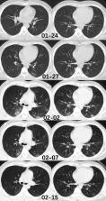
          A total of 85 patients had Hubei travel/residence history, while another 49 had contact with people from Hubei and 15 had no traceable exposure history to Hubei. Fever, cough and expectoration were the most common symptoms, 14 patients had decreased oxygen saturation, 33 had leukopenia, 53 had lymphopenia, and 82 had elevated C-reactive protein. On chest computed tomography (CT), lung segments 6 and 10 were mostly involved. A total of 287 segments presented ground glass opacity, 637 presented mixed opacity and 170 presented consolidation. Lesions were more localized in the peripheral lung with a patchy form. No significant difference was found between patients with or without Hubei exposure history. Seventeen patients had normal CT on admission of these, 12 had negative findings even10 days later.

          Interpretation

          Most patients presented with a mild infection in our study. The imaging pattern of multifocal peripheral ground glass or mixed opacity with predominance in the lower lung is highly suspicious of COVID-19 in the first week of disease onset. Nevetheless, some patients can present with a normal chest finding despite testing positive for COVID-19. Funding: We did not receive any fundings.

          Related collections

          Most cited references13

          • Record: found
          • Abstract: found
          • Article: not found

          CT Imaging Features of 2019 Novel Coronavirus (2019-nCoV)

          In this retrospective case series, chest CT scans of 21 symptomatic patients from China infected with the 2019 novel coronavirus (2019-nCoV) were reviewed, with emphasis on identifying and characterizing the most common findings. Typical CT findings included bilateral pulmonary parenchymal ground-glass and consolidative pulmonary opacities, sometimes with a rounded morphology and a peripheral lung distribution. Notably, lung cavitation, discrete pulmonary nodules, pleural effusions, and lymphadenopathy were absent. Follow-up imaging in a subset of patients during the study time window often demonstrated mild or moderate progression of disease, as manifested by increasing extent and density of lung opacities. © RSNA, 2020
            Bookmark
            • Record: found
            • Abstract: found
            • Article: not found

            Chest CT for Typical 2019-nCoV Pneumonia: Relationship to Negative RT-PCR Testing

            Some patients with positive chest CT findings may present with negative results of real time reverse-transcription–polymerase chain- reaction (RT-PCR) for 2019 novel coronavirus (2019-nCoV). In this report, we present chest CT findings from five patients with 2019-nCoV infection who had initial negative RT-PCR results. All five patients had typical imaging findings, including ground-glass opacity (GGO) (5 patients) and/or mixed GGO and mixed consolidation (2 patients). After isolation for presumed 2019-nCoV pneumonia, all patients were eventually confirmed with 2019-nCoV infection by repeated swab tests. A combination of repeated swab tests and CT scanning may be helpful when for individuals with high clinical suspicion of nCoV infection but negative RT-PCR screening
              Bookmark
              • Record: found
              • Abstract: found
              • Article: not found

              Thin-section CT of severe acute respiratory syndrome: evaluation of 73 patients exposed to or with the disease.

              To retrospectively analyze the thin-section computed tomographic (CT) features in patients with severe acute respiratory syndrome (SARS) at the authors' institution. From March 11, 2003, to April 2, 2003, 74 patients with symptoms and signs suggestive of SARS underwent CT of the thorax; all underwent thin-section CT except for one patient who underwent conventional CT. Group 1 (n = 23) patients had symptoms of SARS in keeping with criteria from the Centers for Disease Control and Prevention and a positive chest radiograph. Group 2 (n = 17) patients had a high clinical suspicion of SARS but a normal radiograph. Group 3 (n = 34) patients had minor symptoms and a normal chest radiograph. The thin-section CT images were analyzed for ground-glass opacification or consolidation, lesion size in each lung segment, peripheral or central location, interstitial thickening, and other abnormalities. Thin-section CT scans were abnormal only for patients in groups 1 and 2. The patient with only conventional CT scans was in group 3; scans for group 3 patients were normal. Affected segments were predominantly in the lower lobes (91 of 149 affected segments). Common findings included ground-glass opacification, sometimes with consolidation, and interlobular septal and intralobular interstitial thickening. The size of each lesion and the total number of segments involved were smaller in group 2 patients. A majority of patients in group 1 (14 of 23) had mixed central and peripheral lesions. In group 2, however, peripheral lesions were more common (10 of 17). In both groups, a purely central lesion was uncommon (one of 23 in group 1 and two of 17 in group 2). Common thin-section CT features of SARS are ground-glass opacification and lower lobe and peripheral distribution. Copyright RSNA, 2003.
                Bookmark

                Author and article information

                Contributors
                Journal
                J Infect
                J. Infect
                The Journal of Infection
                W.B. Saunders
                0163-4453
                1532-2742
                26 February 2020
                26 February 2020
                :
                Affiliations
                [a ]Department of Radiology, Shanghai Jiao Tong University Medical School Affiliated Ruijin Hospital, No.197 Ruijin Er Road, Shanghai 200025, China
                [b ]Department of Radiology, Ruian People's Hospital, Ruian 325200, Zhejiang province, China
                [c ]Department of Radiology, Yueqing People's Hospital, Yueqing 325600, Zhejiang province, China
                [d ]Department of Infectious Diseases, Wenzhou Central Hospital, Wenzhou 325000, Zhejiang province, China
                [e ]Department of Infectious Diseases, Ruian People's Hospital, Ruian 325200, Zhejiang province, China
                [f ]Department of Respiratory and Critical Care Medicine, Shanghai Jiao Tong University Medical School Affiliated Ruijin Hospital, No.197 Ruijin Er Road, Shanghai 200025, China
                Author notes
                [1]

                These authors made equal contributions.

                Article
                S0163-4453(20)30099-2
                10.1016/j.jinf.2020.02.016
                7102539
                32112884
                ec361fd9-cdc1-40c4-af31-2a27b3da89c0
                © 2020 The British Infection Association. Published by Elsevier Ltd. All rights reserved.

                Since January 2020 Elsevier has created a COVID-19 resource centre with free information in English and Mandarin on the novel coronavirus COVID-19. The COVID-19 resource centre is hosted on Elsevier Connect, the company's public news and information website. Elsevier hereby grants permission to make all its COVID-19-related research that is available on the COVID-19 resource centre - including this research content - immediately available in PubMed Central and other publicly funded repositories, such as the WHO COVID database with rights for unrestricted research re-use and analyses in any form or by any means with acknowledgement of the original source. These permissions are granted for free by Elsevier for as long as the COVID-19 resource centre remains active.

                History
                : 21 February 2020
                Categories
                Article

                Infectious disease & Microbiology
                covid-19,infection,outside hubei,ct,lung
                Infectious disease & Microbiology
                covid-19, infection, outside hubei, ct, lung

                Comments

                Comment on this article