1
views
0
recommends
+1 Recommend
0 collections
    0
    shares
      • Record: found
      • Abstract: found
      • Article: found
      Is Open Access

      Recommendations for intervertebral disc notochordal cell investigation: From isolation to characterization

      research-article

      Read this article at

      Bookmark
          There is no author summary for this article yet. Authors can add summaries to their articles on ScienceOpen to make them more accessible to a non-specialist audience.

          Abstract

          Background

          Lineage‐tracing experiments have established that the central region of the mature intervertebral disc, the nucleus pulposus (NP), develops from the embryonic structure called “the notochord”. However, changes in the cells derived from the notochord which form the NP (i.e., notochordal cells [NCs]), in terms of their phenotype and functional identity from early developmental stages to skeletal maturation are less understood. These key issues require further investigation to better comprehend the role of NCs in homeostasis and degeneration as well as their potential for regeneration. Progress in utilizing NCs is currently hampered due to poor consistency and lack of consensus methodology for in vitro NC extraction, manipulation, and characterization.

          Methods

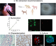
          Here, an international group has come together to provide key recommendations and methodologies for NC isolation within key species, numeration, in vitro manipulation and culture, and characterization.

          Results

          Recommeded protocols are provided for isolation and culture of NCs. Experimental testing provided recommended methodology for numeration of NCs. The issues of cryopreservation are demonstrated, and a pannel of immunohistochemical markers are provided to inform NC characterization.

          Conclusions

          Together we hope this article provides a road map for in vitro studies of NCs to support advances in research into NC physiology and their potential in regenerative therapies.

          Abstract

          To date, there is a lack of consensus for the extraction, numeration, in vitro culture, and characterization of intervertebral disc notochordal cells (NCs). With large variations in methodological approaches affecting progress in this field. Thus, this article aims to provide key recommendations and methodologies for NC isolation, numeration, in vitro manipulation, and characterization to support research into NC physiology and potential in regenerative therapies.

          Related collections

          Most cited references137

          • Record: found
          • Abstract: found
          • Article: not found

          Passaging and colony expansion of human pluripotent stem cells by enzyme-free dissociation in chemically defined culture conditions.

          This protocol describes an EDTA-based passaging procedure to be used with chemically defined E8 medium that serves as a tool for basic and translational research into human pluripotent stem cells (PSCs). In this protocol, passaging one six-well or 10-cm plate of cells takes about 6-7 min. This enzyme-free protocol achieves maximum cell survival without enzyme neutralization, centrifugation or drug treatment. It also allows for higher throughput, requires minimal material and limits contamination. Here we describe how to produce a consistent E8 medium for routine maintenance and reprogramming and how to incorporate the EDTA-based passaging procedure into human induced PSC (iPSC) derivation, colony expansion, cryopreservation and teratoma formation. This protocol has been successful in routine cell expansion, and efficient for expanding large-volume cultures or a large number of cells with preferential dissociation of PSCs. Effective for all culture stages, this procedure provides a consistent and universal approach to passaging human PSCs in E8 medium.
            Bookmark
            • Record: found
            • Abstract: found
            • Article: not found

            The genetic basis of mammalian neurulation.

            More than 80 mutant mouse genes disrupt neurulation and allow an in-depth analysis of the underlying developmental mechanisms. Although many of the genetic mutants have been studied in only rudimentary detail, several molecular pathways can already be identified as crucial for normal neurulation. These include the planar cell-polarity pathway, which is required for the initiation of neural tube closure, and the sonic hedgehog signalling pathway that regulates neural plate bending. Mutant mice also offer an opportunity to unravel the mechanisms by which folic acid prevents neural tube defects, and to develop new therapies for folate-resistant defects.
              Bookmark
              • Record: found
              • Abstract: found
              • Article: not found

              Diversity of intervertebral disc cells: phenotype and function.

              The intervertebral disc (IVD) is a moderately moving joint that is located between the bony vertebrae and provides flexibility and load transmission throughout the spinal column. The disc is composed of different but interrelated tissues, including the central highly hydrated nucleus pulposus (NP), the surrounding elastic and fibrous annulus fibrosus (AF), and the cartilaginous endplate (CEP), which provides the connection to the vertebral bodies. Each of these tissues has a different function and consists of a specific matrix structure that is maintained by a cell population with distinct phenotype. Although the healthy IVD is able to balance the slow matrix turnover of synthesis and degradation, this balance is often disturbed, leading to degenerative disorders. Successful therapeutic management of IVD degeneration requires a profound understanding of the cellular and molecular characteristics of the functional IVD. Hence, the phenotype of IVD cells has been of significant interest from multiple perspectives, including development, growth, remodelling, degeneration and repair. One major challenge that complicates our understanding of the disc cells is that both the cellular phenotype and the extracellular matrix strongly depend on disc maturity and health and as a consequence are continuously evolving. This review delineates the diversity of the cell types found in the intervertebral disc, with emphasis on human, but with reference to other species. The cells of the NP appear rounded and express a proteoglycan-rich matrix, whereas the more elongated AF cells are embedded in a collagen fibre matrix and the CEPs represent a layer of cartilage. Even though all disc cells have often been referred to as 'intervertebral disc chondrocytes', distinct phenotypical differences in comparison with articular chondrocytes exist and have been reported recently. The availability of more specific markers has also improved our understanding of progenitor cell differentiation towards an IVD cell phenotype. Ultimately, new cell- and tissue-engineering approaches to regenerative therapies will only be successful if the specific characteristics of the individual tissues and their context in the function of the whole organ, are taken into consideration. © 2012 The Authors. Journal of Anatomy © 2012 Anatomical Society.
                Bookmark

                Author and article information

                Contributors
                c.lemaitre@sheffield.ac.uk
                Journal
                JOR Spine
                JOR Spine
                10.1002/(ISSN)2572-1143
                JSP2
                JOR Spine
                John Wiley & Sons, Inc. (Hoboken, USA )
                2572-1143
                09 July 2023
                September 2023
                : 6
                : 3 ( doiID: 10.1002/jsp2.v6.3 )
                : e1272
                Affiliations
                [ 1 ] Department of Oncology and Metabolism Medical School, The University of Sheffield Sheffield UK
                [ 2 ] Biomolecular Sciences Research Centre Sheffield Hallam University Sheffield UK
                [ 3 ] Department of Clinical Sciences Faculty of Veterinary Medicine, Utrecht University Utrecht The Netherlands
                [ 4 ] Division of Cell Matrix Biology and Regenerative Medicine, School of Biological Sciences, Faculty of Biology, Medicine and Health The University of Manchester Manchester UK
                [ 5 ] School of Biomedical Sciences The University of Hong Kong Pokfulam Hong Kong China
                [ 6 ] Regenerative Medicine and Skeleton, RMeS Nantes Université, Oniris, CHU Nantes, INSERM, UMR 1229 Nantes France
                [ 7 ] NIHR Manchester Biomedical Research Centre Central Manchester Foundation Trust, Manchester Academic Health Science Centre Manchester UK
                Author notes
                [*] [* ] Correspondence

                Christine L. Le Maitre, Department of Oncology and Metabolism, Medical School, The University of Sheffield, Beech Hill Road, Sheffield S10 2RX, UK.

                Email: c.lemaitre@ 123456sheffield.ac.uk

                Author information
                https://orcid.org/0000-0002-4456-6167
                https://orcid.org/0009-0004-3598-9047
                https://orcid.org/0000-0003-3717-4787
                https://orcid.org/0000-0002-8820-0407
                https://orcid.org/0000-0002-7637-4135
                https://orcid.org/0000-0002-2333-7162
                https://orcid.org/0000-0003-4489-7107
                Article
                JSP21272
                10.1002/jsp2.1272
                10540834
                37780826
                ec2de854-cac4-493b-af2f-bd39e2e639a2
                © 2023 The Authors. JOR Spine published by Wiley Periodicals LLC on behalf of Orthopaedic Research Society.

                This is an open access article under the terms of the http://creativecommons.org/licenses/by-nc/4.0/ License, which permits use, distribution and reproduction in any medium, provided the original work is properly cited and is not used for commercial purposes.

                History
                : 16 June 2023
                : 14 April 2023
                : 21 June 2023
                Page count
                Figures: 12, Tables: 2, Pages: 22, Words: 15091
                Funding
                Funded by: European Union's Horizon 2020
                Funded by: iPSpine
                Award ID: 825925
                Funded by: NC‐CHOICE
                Award ID: 19251
                Funded by: TTW
                Funded by: Dutch Research Council (NWO) , doi 10.13039/501100003246;
                Funded by: Dutch Arthritis Society , doi 10.13039/100018286;
                Award ID: LLP22
                Funded by: RGC European Union – Hong Kong Research and Innovation Cooperation Co‐Funding Mechanism
                Award ID: E‐HKU703/18
                Funded by: Region Pays de la Loire
                Funded by: RFI BIOREGATE (CAVEODISC)
                Funded by: French Society of Rheumatology (Spherodisc)
                Categories
                Research Article
                Research Articles
                Custom metadata
                2.0
                September 2023
                Converter:WILEY_ML3GV2_TO_JATSPMC version:6.3.4 mode:remove_FC converted:29.09.2023

                culture systems,development,intervertebral disc,notochordal cells,nucleus pulposus,tissue‐specific progenitor cells

                Comments

                Comment on this article