3
views
0
recommends
+1 Recommend
0 collections
    0
    shares
      • Record: found
      • Abstract: found
      • Article: not found

      Immunolocalization of Metabolite Transporter Proteins in a Model Cnidarian-Dinoflagellate Symbiosis

      Read this article at

      ScienceOpenPublisherPubMed
      Bookmark
          There is no author summary for this article yet. Authors can add summaries to their articles on ScienceOpen to make them more accessible to a non-specialist audience.

          Abstract

          Coral reefs are in serious decline, in particular due to the thermally induced dysfunction of the cnidarian-dinoflagellate symbiosis that underlies their success. Yet our ability to react to this crisis is hindered by limited knowledge of how this symbiosis functions.

          ABSTRACT

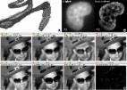
          Bidirectional nutrient flow between partners is integral to the cnidarian-dinoflagellate endosymbiosis. However, our current knowledge of the transporter proteins that regulate nutrient and metabolite trafficking is nascent. Four transmembrane transporters that likely play an important role in interpartner nitrogen and carbon exchange were investigated with immunocytochemistry in the model sea anemone Exaiptasia diaphana (“Aiptasia”; strain NZ1): ammonium transporter 1 (AMT1), V-type proton ATPase (VHA), facilitated glucose transporter member 8 (GLUT8), and aquaporin-3 (AQP3). Anemones lacking symbionts were compared with those in symbiosis with either their typical, homologous dinoflagellate symbiont, Breviolum minutum , or the heterologous species, Durusdinium trenchii and Symbiodinium microadriaticum . AMT1 and VHA were only detected in symbiotic Aiptasia, irrespective of symbiont type. However, GLUT8 and AQP3 were detected in both symbiotic and aposymbiotic states. All transporters were localized to both the epidermis and gastrodermis, though localization patterns in host tissues were heavily influenced by symbiont identity, with S. microadriaticum -colonized anemones showing the most distinct patterns. These patterns suggested disruption of fixed carbon and inorganic nitrogen fluxes when in symbiosis with heterologous versus homologous symbionts. This study enhances our understanding of nutrient transport and host-symbiont integration, while providing a platform for further investigation of nutrient transporters and the host-symbiont interface in the cnidarian-dinoflagellate symbiosis.

          IMPORTANCE Coral reefs are in serious decline, in particular due to the thermally induced dysfunction of the cnidarian-dinoflagellate symbiosis that underlies their success. Yet our ability to react to this crisis is hindered by limited knowledge of how this symbiosis functions. Indeed, we still have much to learn about the cellular integration that determines whether a particular host-symbiont combination can persist, and hence whether corals might be able to adapt by acquiring new, more thermally resistant symbionts. Here, we employed immunocytochemistry to localize and quantify key nutrient transporters in tissues of the sea anemone Aiptasia, a globally adopted model system for this symbiosis, and compared the expression of these transporters when the host is colonized by native versus nonnative symbionts. We showed a clear link between transporter expression and symbiont identity, elucidating the cellular events that dictate symbiosis success, and we provide a methodological platform for further examination of cellular integration in this ecologically important symbiosis.

          Related collections

          Most cited references104

          • Record: found
          • Abstract: found
          • Article: not found

          Fiji: an open-source platform for biological-image analysis.

          Fiji is a distribution of the popular open-source software ImageJ focused on biological-image analysis. Fiji uses modern software engineering practices to combine powerful software libraries with a broad range of scripting languages to enable rapid prototyping of image-processing algorithms. Fiji facilitates the transformation of new algorithms into ImageJ plugins that can be shared with end users through an integrated update system. We propose Fiji as a platform for productive collaboration between computer science and biology research communities.
            Bookmark
            • Record: found
            • Abstract: found
            • Article: found
            Is Open Access

            ImageJ2: ImageJ for the next generation of scientific image data

            Background ImageJ is an image analysis program extensively used in the biological sciences and beyond. Due to its ease of use, recordable macro language, and extensible plug-in architecture, ImageJ enjoys contributions from non-programmers, amateur programmers, and professional developers alike. Enabling such a diversity of contributors has resulted in a large community that spans the biological and physical sciences. However, a rapidly growing user base, diverging plugin suites, and technical limitations have revealed a clear need for a concerted software engineering effort to support emerging imaging paradigms, to ensure the software’s ability to handle the requirements of modern science. Results We rewrote the entire ImageJ codebase, engineering a redesigned plugin mechanism intended to facilitate extensibility at every level, with the goal of creating a more powerful tool that continues to serve the existing community while addressing a wider range of scientific requirements. This next-generation ImageJ, called “ImageJ2” in places where the distinction matters, provides a host of new functionality. It separates concerns, fully decoupling the data model from the user interface. It emphasizes integration with external applications to maximize interoperability. Its robust new plugin framework allows everything from image formats, to scripting languages, to visualization to be extended by the community. The redesigned data model supports arbitrarily large, N-dimensional datasets, which are increasingly common in modern image acquisition. Despite the scope of these changes, backwards compatibility is maintained such that this new functionality can be seamlessly integrated with the classic ImageJ interface, allowing users and developers to migrate to these new methods at their own pace. Conclusions Scientific imaging benefits from open-source programs that advance new method development and deployment to a diverse audience. ImageJ has continuously evolved with this idea in mind; however, new and emerging scientific requirements have posed corresponding challenges for ImageJ’s development. The described improvements provide a framework engineered for flexibility, intended to support these requirements as well as accommodate future needs. Future efforts will focus on implementing new algorithms in this framework and expanding collaborations with other popular scientific software suites. Electronic supplementary material The online version of this article (doi:10.1186/s12859-017-1934-z) contains supplementary material, which is available to authorized users.
              Bookmark
              • Record: found
              • Abstract: found
              • Article: not found

              A guided tour into subcellular colocalization analysis in light microscopy.

              It is generally accepted that the functional compartmentalization of eukaryotic cells is reflected by the differential occurrence of proteins in their compartments. The location and physiological function of a protein are closely related; local information of a protein is thus crucial to understanding its role in biological processes. The visualization of proteins residing on intracellular structures by fluorescence microscopy has become a routine approach in cell biology and is increasingly used to assess their colocalization with well-characterized markers. However, image-analysis methods for colocalization studies are a field of contention and enigma. We have therefore undertaken to review the most currently used colocalization analysis methods, introducing the basic optical concepts important for image acquisition and subsequent analysis. We provide a summary of practical tips for image acquisition and treatment that should precede proper colocalization analysis. Furthermore, we discuss the application and feasibility of colocalization tools for various biological colocalization situations and discuss their respective strengths and weaknesses. We have created a novel toolbox for subcellular colocalization analysis under ImageJ, named JACoP, that integrates current global statistic methods and a novel object-based approach.
                Bookmark

                Author and article information

                Contributors
                (View ORCID Profile)
                (View ORCID Profile)
                (View ORCID Profile)
                (View ORCID Profile)
                (View ORCID Profile)
                Journal
                Applied and Environmental Microbiology
                Appl Environ Microbiol
                American Society for Microbiology
                0099-2240
                1098-5336
                June 28 2022
                June 28 2022
                : 88
                : 12
                Affiliations
                [1 ]School of Biological Sciences, Victoria University of Wellington, Wellington, New Zealand
                [2 ]Department of Plant Biology, The Carnegie Institution for Science, Stanford, California, USA
                [3 ]Department of Integrative Biology, Oregon State University, Corvallis, Oregon, USA
                Article
                10.1128/aem.00412-22
                35678605
                e8aad426-ffd8-4db7-aecb-e12533144e6f
                © 2022

                https://doi.org/10.1128/ASMCopyrightv2

                https://journals.asm.org/non-commercial-tdm-license

                History

                Comments

                Comment on this article