0
views
0
recommends
+1 Recommend
0 collections
    0
    shares
      • Record: found
      • Abstract: found
      • Article: found
      Is Open Access

      Gut-associated lymphoid tissue: a microbiota-driven hub of B cell immunity

      review-article

      Read this article at

      Bookmark
          There is no author summary for this article yet. Authors can add summaries to their articles on ScienceOpen to make them more accessible to a non-specialist audience.

          Highlights

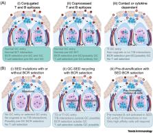
          • Mammalian gut-associated lymphoid tissue (GALT) is chronically activated by the intestinal microbiota throughout life.

          • GALT propagates and selects B cells in germinal centers, including B cells recognizing T-cell-independent carbohydrate antigens.

          • GALT supports the development of systemic B cells in the so-called GALT species, including humans.

          • In humans, GALT supports the development of innate-like marginal zone B cells that circulate in blood, mostly reside in the spleen, and can protect the lungs.

          Significance

          Gut-associated lymphoid tissue (GALT) is located throughout the gastrointestinal tract and is indispensable for the maintenance of gut homeostasis and human health. Recent advances increase our understanding of B cell responses to microbiota in GALT that can generate antibodies to antigens often shared by microbiota species, including T cell independent, yet germinal-center-matured responses. Indeed, chronic stimulation of GALT B cells generates a niche for B cell maturation and propagation in some species, including marginal zone B cells in humans.

          Abstract

          The diverse gut microbiota, which is associated with mucosal health and general wellbeing, maintains gut-associated lymphoid tissues (GALT) in a chronically activated state, including sustainment of germinal centers in a context of high antigenic load. This influences the rules for B cell engagement with antigen and the potential consequences. Recent data have highlighted differences between GALT and other lymphoid tissues. For example, GALT propagates IgA responses against glycans that show signs of having been generated in germinal centers. Other findings suggest that humans are among those species where GALT supports the diversification, propagation, and possibly selection of systemic B cells. Here, we review novel findings that identify GALT as distinctive, and able to support these processes.

          Related collections

          Most cited references84

          • Record: found
          • Abstract: found
          • Article: not found

          Visualization and analysis of gene expression in tissue sections by spatial transcriptomics.

          Analysis of the pattern of proteins or messengerRNAs (mRNAs) in histological tissue sections is a cornerstone in biomedical research and diagnostics. This typically involves the visualization of a few proteins or expressed genes at a time. We have devised a strategy, which we call "spatial transcriptomics," that allows visualization and quantitative analysis of the transcriptome with spatial resolution in individual tissue sections. By positioning histological sections on arrayed reverse transcription primers with unique positional barcodes, we demonstrate high-quality RNA-sequencing data with maintained two-dimensional positional information from the mouse brain and human breast cancer. Spatial transcriptomics provides quantitative gene expression data and visualization of the distribution of mRNAs within tissue sections and enables novel types of bioinformatics analyses, valuable in research and diagnostics.
            Bookmark
            • Record: found
            • Abstract: found
            • Article: not found

            Distinct Effector B Cells Induced by Unregulated Toll-like Receptor 7 Contribute to Pathogenic Responses in Systemic Lupus Erythematosus

            Systemic Lupus Erythematosus (SLE) is characterized by B-cells lacking IgD and CD27 (double negative; DN). We show that DN cell expansions reflected a subset of CXCR5 − CD11c + cells (DN2) representing pre-plasma cells (PC). DN2 cells predominated in African-American patients with active disease and nephritis, anti-Smith and anti-RNA autoantibodies. They expressed a T-bet transcriptional network; increased toll-like receptor-7 (TLR7); lacked the negative TLR regulator TRAF5; and were hyper-responsive to TLR7. DN2 cells shared with activated naïve cells (aNAV), phenotypic and functional features, and similar transcriptomes. Their PC differentiation and autoantibody production was driven by TLR7 in an interleukin-21 (IL-21)-mediated fashion. An in vivo developmental link between aNAV, DN2 cells and PC was demonstrated by clonal sharing. This study defines a distinct differentiation fate of autoreactive naïve B cells into PC precursors with hyper-responsiveness to innate stimuli, as well as establishes prominence of extra-follicular B-cell activation in SLE, and identifies therapeutic targets. The role of extrafollicular B cells in human systemic lupus is unknown. Jenks et al . define the main components of this pathway and its prominence in severe disease. Its activation is mediated by hyper-responsiveness to toll-like receptor-7 and leads to the generation of autoreactive antibody-secreting plasmablasts.
              Bookmark
              • Record: found
              • Abstract: found
              • Article: not found

              Uptake through glycoprotein 2 of FimH(+) bacteria by M cells initiates mucosal immune response.

              The mucosal immune system forms the largest part of the entire immune system, containing about three-quarters of all lymphocytes and producing grams of secretory IgA daily to protect the mucosal surface from pathogens. To evoke the mucosal immune response, antigens on the mucosal surface must be transported across the epithelial barrier into organized lymphoid structures such as Peyer's patches. This function, called antigen transcytosis, is mediated by specialized epithelial M cells. The molecular mechanisms promoting this antigen uptake, however, are largely unknown. Here we report that glycoprotein 2 (GP2), specifically expressed on the apical plasma membrane of M cells among enterocytes, serves as a transcytotic receptor for mucosal antigens. Recombinant GP2 protein selectively bound a subset of commensal and pathogenic enterobacteria, including Escherichia coli and Salmonella enterica serovar Typhimurium (S. Typhimurium), by recognizing FimH, a component of type I pili on the bacterial outer membrane. Consistently, these bacteria were colocalized with endogenous GP2 on the apical plasma membrane as well as in cytoplasmic vesicles in M cells. Moreover, deficiency of bacterial FimH or host GP2 led to defects in transcytosis of type-I-piliated bacteria through M cells, resulting in an attenuation of antigen-specific immune responses in Peyer's patches. GP2 is therefore a previously unrecognized transcytotic receptor on M cells for type-I-piliated bacteria and is a prerequisite for the mucosal immune response to these bacteria. Given that M cells are considered a promising target for oral vaccination against various infectious diseases, the GP2-dependent transcytotic pathway could provide a new target for the development of M-cell-targeted mucosal vaccines.
                Bookmark

                Author and article information

                Contributors
                Journal
                Trends Immunol
                Trends Immunol
                Trends in Immunology
                Elsevier Science Ltd
                1471-4906
                1471-4981
                1 March 2024
                March 2024
                : 45
                : 3
                : 211-223
                Affiliations
                [1 ]Department of Translational Medicine – Human Immunology, Lund University, J Waldenströms gata 35, Malmö, Sweden
                [2 ]Department of Clinical Immunology and Transfusion Medicine, Sahlgrenska University Hospital, Region Västra Götaland, Gothenburg, Sweden
                [3 ]Peter Gorer Department of Immunobiology, King’s College London, Guy’s Hospital Campus, St Thomas’ Street, London SE1 9RT, UK
                Author notes
                Article
                S1471-4906(24)00006-1
                10.1016/j.it.2024.01.006
                11227984
                38402045
                e70bc15c-795a-474e-b379-ed4714fa987f
                © 2024 The Author(s)

                This is an open access article under the CC BY license (http://creativecommons.org/licenses/by/4.0/).

                History
                Categories
                Review

                Immunology
                Immunology

                Comments

                Comment on this article