23
views
0
recommends
+1 Recommend
0 collections
    0
    shares
      • Record: found
      • Abstract: found
      • Article: found
      Is Open Access

      Safety of bioabsorbable implants in vitro

      research-article

      Read this article at

      Bookmark
          There is no author summary for this article yet. Authors can add summaries to their articles on ScienceOpen to make them more accessible to a non-specialist audience.

          Abstract

          Background

          The aim of the present study was to investigate the safety of bioabsorbable plates and screws in humans.

          Methods

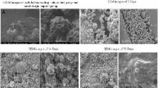
          For this purpose, an implant system based on [poly(lactic-co-glycolic acids)(85:15)] was designed. The system was tested for pH, temperature, and swelling and then its surface morphology was analyzed for surface porosity using environmental electron microscopy. Then, the effects of this bioabsorbable system on the viability and profileration of osteocytes were examined on a molecular level via in vitro experiments. A [poly(lactic-co-glycolic acids)(90:10)] bioabsorbable implant, which is commercially available and used in orthopedic surgery, was used as control group. For the statistical evaluation of the data obtained in the present study, the groups were compared by Tukey HSD test following ANOVA. The significance level was set as p < 0.05.

          Results

          It was observed that the osteocytes cultivated on the PLGA system designed in the present study included more live cells and allowed more proliferation compared to the control.

          Conclusion

          One of the criteria in the selection of implants for orthopedic surgery is that a good implant should not need removal and thus a second surgery. In the present study, a bioabsorbable implant was designed considering this criterion. The present study is the first step to prove the safety of this new design by in vitro toxicity and viability experiments.

          Related collections

          Most cited references39

          • Record: found
          • Abstract: found
          • Article: not found

          Making tissue engineering scaffolds work. Review: the application of solid freeform fabrication technology to the production of tissue engineering scaffolds.

          Tissue engineering is a new and exciting technique which has the potential to create tissues and organs de novo. It involves the in vitro seeding and attachment of human cells onto a scaffold. These cells then proliferate, migrate and differentiate into the specific tissue while secreting the extracellular matrix components required to create the tissue. It is evident, therefore, that the choice of scaffold is crucial to enable the cells to behave in the required manner to produce tissues and organs of the desired shape and size. Current scaffolds, made by conventional scaffold fabrication techniques, are generally foams of synthetic polymers. The cells do not necessarily recognise such surfaces, and most importantly cells cannot migrate more than 500 microm from the surface. The lack of oxygen and nutrient supply governs this depth. Solid freeform fabrication (SFF) uses layer-manufacturing strategies to create physical objects directly from computer-generated models. It can improve current scaffold design by controlling scaffold parameters such as pore size, porosity and pore distribution, as well as incorporating an artificial vascular system, thereby increasing the mass transport of oxygen and nutrients into the interior of the scaffold and supporting cellular growth in that region. Several SFF systems have produced tissue engineering scaffolds with this concept in mind which will be the main focus of this review. We are developing scaffolds from collagen and with an internal vascular architecture using SFF. Collagen has major advantages as it provides a favourable surface for cellular attachment. The vascular system allows for the supply of nutrients and oxygen throughout the scaffold. The future of tissue engineering scaffolds is intertwined with SFF technologies.
            Bookmark
            • Record: found
            • Abstract: found
            • Article: not found

            Understanding the biodegradation of polyurethanes: from classical implants to tissue engineering materials.

            After almost half a century of use in the health field, polyurethanes (PUs) remain one of the most popular group of biomaterials applied for medical devices. Their popularity has been sustained as a direct result of their segmented block copolymeric character, which endows them with a wide range of versatility in terms of tailoring their physical properties, blood and tissue compatibility, and more recently their biodegradation character. While they became recognized in the 1970s and 1980s as the blood contacting material of choice in a wide range of cardiovascular devices their application in long-term implants fell under scrutiny with the failure of pacemaker leads and breast implant coatings containing PUs in the late 1980s. During the next decade PUs became extensively researched for their relative sensitivity to biodegradation and the desire to further understand the biological mechanisms for in vivo biodegradation. The advent of molecular biology into mainstream biomedical engineering permitted the probing of molecular pathways leading to the biodegradation of these materials. Knowledge gained throughout the 1990s has not only yielded novel PUs that contribute to the enhancement of biostability for in vivo long-term applications, but has also been translated to form a new class of bioresorbable materials with all the versatility of PUs in terms of physical properties but now with a more integrative nature in terms of biocompatibility. The current review will briefly survey the literature, which initially identified the problem of PU degradation in vivo and the subsequent studies that have led to the field's further understanding of the biological processes mediating the breakdown. An overview of research emerging on PUs sought for use in combination (drug + polymer) products and tissue regeneration applications will then be presented.
              Bookmark
              • Record: found
              • Abstract: found
              • Article: not found

              Characterization of porous collagen/hyaluronic acid scaffold modified by 1-ethyl-3-(3-dimethylaminopropyl)carbodiimide cross-linking.

              In order to develop a scaffolding material for tissue regeneration, porous matrices containing collagen and hyaluronic acid were fabricated by freeze drying at -20 degrees C, -70 degrees C or -196 degrees C. The fabricated porous membranes were cross-linked using 1-ethyl-3-(3-dimethyl aminopropyl) carbodiimide (EDC) in a range of 1-100 mM concentrations for enhancing mechanical stability of the composite matrix. Scanning electron microscope (SEM) views of the matrices demonstrated that the matrices obtained before cross-linking process had interconnected pores with mean diameters of 40, 90 or 230 microm and porosity of 58-66% according to the freezing temperature, and also the porous structures after cross-linking process were retained. The swelling test and IR spectroscopic measurement of different cross-linked membranes were carried out as a measure of the extent of cross-linking. The swelling behavior of cross-linked membranes showed no significant differences as cross-linking degree increased. FT-IR spectra showed the increase of the intensity of the absorbencies at amide bonds (1655, 1546, 1458 cm(-1)) compared to that of CH bond (2930 cm(-1)). In enzymatic degradation test, EDC treated membranes showed significant enhancement of the resistance to collagenase activity in comparison with 0.625% glutaraldehyde treated membranes. In cytotoxicity test using L929 fibroblastic cells, the EDC-cross-linked membranes demonstrated no significant toxicity.
                Bookmark

                Author and article information

                Contributors
                +9053 3722 7971 , misyar2003@yahoo.com
                ibrahimyilmaz77@yahoo.com
                gnusran@hotmail.com
                olcayguler77@gmail.com
                seralple@hotmail.com
                mahirogullari@yahoo.com
                Journal
                BMC Surg
                BMC Surg
                BMC Surgery
                BioMed Central (London )
                1471-2482
                12 December 2015
                12 December 2015
                2015
                : 15
                : 127
                Affiliations
                [ ]Department of Orthopaedic and Traumatology, Istanbul Medipol University School of Medicine, Bagcilar, 34214 Istanbul, Turkey
                [ ]Department of Pharmacovigilance and Rational Drug Use Team, Pharmacologist Pharmacist, M.Sc. Republic of Turkey, Ministry of Health, State Hospital, 59100 Tekirdag, Turkey
                [ ]Department of Orthopaedic and Traumatology, Canakkale Onsekizmart University School of Medicine, 17000 Canakkale, Turkey
                Article
                111
                10.1186/s12893-015-0111-4
                4676853
                e3f4ac04-b4ba-4884-967c-11dff1361c64
                © Isyar et al. 2015

                Open AccessThis article is distributed under the terms of the Creative Commons Attribution 4.0 International License ( http://creativecommons.org/licenses/by/4.0/), which permits unrestricted use, distribution, and reproduction in any medium, provided you give appropriate credit to the original author(s) and the source, provide a link to the Creative Commons license, and indicate if changes were made. The Creative Commons Public Domain Dedication waiver ( http://creativecommons.org/publicdomain/zero/1.0/) applies to the data made available in this article, unless otherwise stated.

                History
                : 22 August 2015
                : 1 December 2015
                Categories
                Research Article
                Custom metadata
                © The Author(s) 2015

                Surgery
                bioabsorbable implant,osteocyte proliferation,osteoblastic activity,poly(lactic-co-glycolic acids)

                Comments

                Comment on this article