15
views
0
recommends
+1 Recommend
1 collections
    0
    shares

      To submit to Bentham Journals, please click here

      • Record: found
      • Abstract: found
      • Article: found
      Is Open Access

      Comparison of Mesiodistal Root Angulation Measured from Conventional and CBCT Derived Panoramic Radiographs in Orthodontic Patients

      research-article

      Read this article at

      Bookmark
          There is no author summary for this article yet. Authors can add summaries to their articles on ScienceOpen to make them more accessible to a non-specialist audience.

          Abstract

          Introduction:

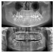
          Use of cone beam computed tomography (CBCT) in orthodontics is increasing; however, some patients started treatment with conventional images. The objective of this study is to manipulate CBCT panoramic reconstruction to make it comparable to conventional panoramic image and to compare mesiodistal root angulations on both images.

          Materials and Methods:

          Concurrent conventional panoramics and CBCT volumes were obtained from 40 subjects. CBCT volumes were manipulated to generate pan-like images that mimic the occlusal plane angle of the corresponding panoramic, allowing comparison of mesiodistal root angulations and determination of the head-tilt required to produce the reconstruction.

          Results:

          Clinically meaningful differences ( p < .05) in the mesiodistal root angulations between standard panoramics and CBCT reconstructions emerged for 13 out of 24 teeth (54%). Greatest variations were seen in the maxillary and mandibular sextants and in first molar regions. Ideal axial head-tilt for image acquisition was determined to be with Frankfort horizontal plane 3.3 o nose down.

          Conclusion:

          CBCT pan images must be used with caution due to variation between methods in specific areas of arches. The images can be useful for the assessment of mesiodistal root angulations if the volume is properly manipulated to create a pan-like image.

          Related collections

          Most cited references21

          • Record: found
          • Abstract: not found
          • Article: not found

          The six keys to normal occlusion.

            Bookmark
            • Record: found
            • Abstract: found
            • Article: not found

            Radiographic examination of the temporomandibular joint using cone beam computed tomography.

            Cone beam CT (CBCT) is a new technique for maxillofacial imaging. We describe a reconstruction technique for radiographic examination of the temporomandibular joint (TMJ) using CBCT, and we further present four cases where the technique was employed. The technique provides a complete radiographic investigation of the bony components of the TMJ. The reconstructed images are of high diagnostic quality. The examination time is shorter and the patient dose is lower than that with conventional CT. It may therefore be considered as the imaging technique of choice when investigation of bony changes of the TMJ is the task at hand.
              Bookmark
              • Record: found
              • Abstract: found
              • Article: not found

              Comparison of mesiodistal root angulation with posttreatment panoramic radiographs and cone-beam computed tomography.

              Orthodontists assess mesiodistal root angulations before, during, and after orthodontic treatment as an aid in establishing proper root position. Panoramic imaging has been useful for this purpose and is a valuable screening tool in diagnosis and planning treatment of orthodontic patients. Cone-beam computed tomography (CBCT) for imaging of the craniofacial complex creates the opportunity to evaluate 3-dimensional images compared with traditional 2-dimensional images. The purpose of this project was to compare mesiodistal root angulations by using posttreatment panoramic radiographic images and CBCT scans.
                Bookmark

                Author and article information

                Journal
                Open Dent J
                Open Dent J
                TODENTJ
                The Open Dentistry Journal
                Bentham Open
                1874-2106
                30 June 2017
                2017
                : 11
                : 338-349
                Affiliations
                [1 ]Department of DentoMaxillofacial Radiology and Imaging School of Dentistry, Lebanese University, Beirut, Lebanon
                [2 ]Department of developmental dentistry The University of Texas Health Science Center, San Antonio School of Dentistry, San Antonio, TX, USA
                [3 ]Oral and Maxillofacial Radiology The University of Texas Health Science Center, San Antonio School of Dentistry, San Antonio, TX, USA
                Author notes
                [* ]Address correspondence to this author at the Department of DentoMaxillofacial Radiology and Imaging, School of Dentistry, Lebanese University, Beirut, Lebanon, Tel: 9613302232; E-mails: ibrahim.nasseh@ 123456gmail.com ; @ 123456inasseh.ul.edu.lb
                Article
                TODENTJ-11-338
                10.2174/1874210601711010338
                5543660
                e383ede2-277b-447b-b1a2-7dde3d2e34a8
                © 2017 Nasseh et al.

                This is an open access article distributed under the terms of the Creative Commons Attribution 4.0 International Public License (CC-BY 4.0), a copy of which is available at: ( https://creativecommons.org/licenses/by/4.0/legalcode). This license permits unrestricted use, distribution, and reproduction in any medium, provided the original author and source are credited.

                History
                : 30 November 2016
                : 07 December 2016
                : 14 December 2016
                Categories
                Article
                Suppl-1, M2

                Dentistry
                cbct,mesiodistal root,panoramic radiographs,conventional images,pan-like images
                Dentistry
                cbct, mesiodistal root, panoramic radiographs, conventional images, pan-like images

                Comments

                Comment on this article