25
views
0
recommends
+1 Recommend
0 collections
    0
    shares
      • Record: found
      • Abstract: found
      • Article: found
      Is Open Access

      The BH3-only protein Bik/Blk/Nbk inhibits nuclear translocation of activated ERK1/2 to mediate IFNγ-induced cell death

      research-article

      Read this article at

      Bookmark
          There is no author summary for this article yet. Authors can add summaries to their articles on ScienceOpen to make them more accessible to a non-specialist audience.

          Abstract

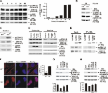
          IFNγ induces cell death in epithelial cells, but the mediator for this death pathway has not been identified. In this study, we find that expression of Bik/Blk/Nbk is increased in human airway epithelial cells (AECs [HAECs]) in response to IFNγ. Expression of Bik but not mutant BikL61G induces and loss of Bik suppresses IFNγ-induced cell death in HAECs. IFNγ treatment and Bik expression increase cathepsin B and D messenger RNA levels and reduce levels of phospho–extracellular regulated kinase 1/2 (ERK1/2) in the nuclei of bik +/+ compared with bik −/− murine AECs. Bik but not BikL61G interacts with and suppresses nuclear translocation of phospho-ERK1/2, and suppression of ERK1/2 activation inhibits IFNγ- and Bik-induced cell death. Furthermore, after prolonged exposure to allergen, hyperplastic epithelial cells persist longer, and nuclear phospho-ERK is more prevalent in airways of IFNγ −/− or bik −/− compared with wild-type mice. These results demonstrate that IFNγ requires Bik to suppress nuclear localization of phospho-ERK1/2 to channel cell death in AECs.

          Related collections

          Most cited references62

          • Record: found
          • Abstract: found
          • Article: not found

          Endoplasmic reticulum stress: cell life and death decisions.

          C. Xu (2005)
          Disturbances in the normal functions of the ER lead to an evolutionarily conserved cell stress response, the unfolded protein response, which is aimed initially at compensating for damage but can eventually trigger cell death if ER dysfunction is severe or prolonged. The mechanisms by which ER stress leads to cell death remain enigmatic, with multiple potential participants described but little clarity about which specific death effectors dominate in particular cellular contexts. Important roles for ER-initiated cell death pathways have been recognized for several diseases, including hypoxia, ischemia/reperfusion injury, neurodegeneration, heart disease, and diabetes.
            Bookmark
            • Record: found
            • Abstract: found
            • Article: not found

            Noxa, a BH3-only member of the Bcl-2 family and candidate mediator of p53-induced apoptosis.

            A critical function of tumor suppressor p53 is the induction of apoptosis in cells exposed to noxious stresses. We report a previously unidentified pro-apoptotic gene, Noxa. Expression of Noxa induction in primary mouse cells exposed to x-ray irradiation was dependent on p53. Noxa encodes a Bcl-2 homology 3 (BH3)-only member of the Bcl-2 family of proteins; this member contains the BH3 region but not other BH domains. When ectopically expressed, Noxa underwent BH3 motif-dependent localization to mitochondria and interacted with anti-apoptotic Bcl-2 family members, resulting in the activation of caspase-9. We also demonstrate that blocking the endogenous Noxa induction results in the suppression of apoptosis. Noxa may thus represent a mediator of p53-dependent apoptosis.
              Bookmark
              • Record: found
              • Abstract: found
              • Article: not found

              A novel assay for apoptosis. Flow cytometric detection of phosphatidylserine expression on early apoptotic cells using fluorescein labelled Annexin V.

              In the early stages of apoptosis changes occur at the cell surface, which until now have remained difficult to recognize. One of these plasma membrane alterations is the translocation of phosphatidylserine (PS) from the inner side of the plasma membrane to the outer layer, by which PS becomes exposed at the external surface of the cell. Annexin V is a Ca2+ dependent phospholipid-binding protein with high affinity for PS. Hence this protein can be used as a sensitive probe for PS exposure upon the cell membrane. Translocation of PS to the external cell surface is not unique to apoptosis, but occurs also during cell necrosis. The difference between these two forms of cell death is that during the initial stages of apoptosis the cell membrane remains intact, while at the very moment that necrosis occurs the cell membrane looses its integrity and becomes leaky. Therefore the measurement of Annexin V binding to the cell surface as indicative for apoptosis has to be performed in conjunction with a dye exclusion test to establish integrity of the cell membrane. This paper describes the results of such an assay, as obtained in cultured HSB-2 cells, rendered apoptotic by irradiation and in human lymphocytes, following dexamethasone treatment. Untreated and treated cells were evaluated for apoptosis by light microscopy, by measuring the amount of hypo-diploid cells using of DNA flow cytometry (FCM) and by DNA electrophoresis to establish whether or not DNA fragmentation had occurred. Annexin V binding was assessed using bivariate FCM, and cell staining was evaluated with fluorescein isothiocyanate (FITC)-labelled Annexin V (green fluorescence), simultaneously with dye exclusion of propidium iodide (PI) (negative for red fluorescence). The test described, discriminates intact cells (FITC-/PI-), apoptotic cells (FITC+/PI-) and necrotic cells (FITC+/PI+). In comparison with existing traditional tests the Annexin V assay is sensitive and easy to perform. The Annexin V assay offers the possibility of detecting early phases of apoptosis before the loss of cell membrane integrity and permits measurements of the kinetics of apoptotic death in relation to the cell cycle. More extensive FCM will allow discrimination between different cell subpopulations, that may or may not be involved in the apoptotic process.
                Bookmark

                Author and article information

                Journal
                J Cell Biol
                jcb
                The Journal of Cell Biology
                The Rockefeller University Press
                0021-9525
                1540-8140
                3 November 2008
                : 183
                : 3
                : 429-439
                Affiliations
                [1 ]Lovelace Respiratory Research Institute, Albuquerque, NM 87108
                [2 ]Department of Pulmonary Medicine, M.D. Anderson Cancer Center, Houston, TX 77030
                Author notes

                Correspondence to Y. Tesfaigzi: ytesfaig@ 123456lrri.org

                Article
                200801186
                10.1083/jcb.200801186
                2575785
                18981230
                e290fb48-4551-46a3-93cd-3ad853213ab8
                © 2008 Mebratu et al.

                This article is distributed under the terms of an Attribution–Noncommercial–Share Alike–No Mirror Sites license for the first six months after the publication date (see http://www.jcb.org/misc/terms.shtml). After six months it is available under a Creative Commons License (Attribution–Noncommercial–Share Alike 3.0 Unported license, as described at http://creativecommons.org/licenses/by-nc-sa/3.0/).

                History
                : 30 January 2008
                : 1 October 2008
                Categories
                Research Articles
                Article

                Cell biology
                Cell biology

                Comments

                Comment on this article