- Record: found
- Abstract: found
- Article: found
Three cases of intra‐abdominal free air onset associated with COPD treated conservatively

Read this article at
Abstract
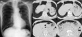
Pneumoperitoneum is caused by respiratory disease in rare cases and can be treated conservatively. It is important to confirm physical abdominal examinations, laboratory data, and radiological findings to avoid unnecessary surgical procedures. The diagnosis of pneumoperitoneum associated with respiratory disease requires the exclusion of other fatal illnesses, especially gastrointestinal perforation.
Abstract
Pneumoperitoneum is caused by respiratory disease in rare cases and can be treated conservatively. It is important to confirm physical abdominal examinations, laboratory data, and radiological findings to avoid unnecessary surgical procedures. The diagnosis of pneumoperitoneum associated with respiratory disease requires the exclusion of other fatal illnesses, especially gastrointestinal perforation.
Related collections
Most cited references11
- Record: found
- Abstract: found
- Article: not found
Computed tomography in pulmonary emphysema.
- Record: found
- Abstract: not found
- Article: not found
Pneumatosis intestinalis: a new concept.
- Record: found
- Abstract: found
- Article: found