5
views
0
recommends
+1 Recommend
0 collections
    0
    shares
      • Record: found
      • Abstract: found
      • Article: found
      Is Open Access

      Effective combinatorial immunotherapy for penile squamous cell carcinoma

      research-article

      Read this article at

      Bookmark
          There is no author summary for this article yet. Authors can add summaries to their articles on ScienceOpen to make them more accessible to a non-specialist audience.

          Abstract

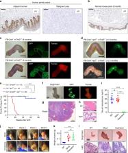
          Penile squamous cell carcinoma (PSCC) accounts for over 95% of penile malignancies and causes significant mortality and morbidity in developing countries. Molecular mechanisms and therapies of PSCC are understudied, owing to scarcity of laboratory models. Herein, we describe a genetically engineered mouse model of PSCC, by co-deletion of Smad4 and Apc in the androgen-responsive epithelium of the penis. Mouse PSCC fosters an immunosuppressive microenvironment with myeloid-derived suppressor cells (MDSCs) as a dominant population. Preclinical trials in the model demonstrate synergistic efficacy of immune checkpoint blockade with the MDSC-diminishing drugs cabozantinib or celecoxib. A critical clinical problem of PSCC is chemoresistance to cisplatin, which is induced by Pten deficiency on the backdrop of Smad4/Apc co-deletion. Drug screen studies informed by targeted proteomics identify a few potential therapeutic strategies for PSCC. Our studies have established what we believe to be essential resources for studying PSCC biology and developing therapeutic strategies.

          Abstract

          Penile squamous cell carcinoma (PSCC) is a cancer that is associated with significant mortality. Here, the authors develop a mouse model of PSCC by co-deletion of Smad4 and Apc in the androgen-responsive penile epithelium, and show synergistic efficacy of checkpoint therapy with cabozantinib or celecoxib in their model.

          Related collections

          Most cited references47

          • Record: found
          • Abstract: found
          • Article: not found

          SOX2 controls tumour initiation and cancer stem-cell functions in squamous-cell carcinoma.

          Cancer stem cells (CSCs) have been reported in various cancers, including in skin squamous-cell carcinoma (SCC). The molecular mechanisms regulating tumour initiation and stemness are still poorly characterized. Here we find that Sox2, a transcription factor expressed in various types of embryonic and adult stem cells, was the most upregulated transcription factor in the CSCs of squamous skin tumours in mice. SOX2 is absent in normal epidermis but begins to be expressed in the vast majority of mouse and human pre-neoplastic skin tumours, and continues to be expressed in a heterogeneous manner in invasive mouse and human SCCs. In contrast to other SCCs, in which SOX2 is frequently genetically amplified, the expression of SOX2 in mouse and human skin SCCs is transcriptionally regulated. Conditional deletion of Sox2 in the mouse epidermis markedly decreases skin tumour formation after chemical-induced carcinogenesis. Using green fluorescent protein (GFP) as a reporter of Sox2 transcriptional expression (SOX2-GFP knock-in mice), we showed that SOX2-expressing cells in invasive SCC are greatly enriched in tumour-propagating cells, which further increase upon serial transplantations. Lineage ablation of SOX2-expressing cells within primary benign and malignant SCCs leads to tumour regression, consistent with the critical role of SOX2-expressing cells in tumour maintenance. Conditional Sox2 deletion in pre-existing skin papilloma and SCC leads to tumour regression and decreases the ability of cancer cells to be propagated upon transplantation into immunodeficient mice, supporting the essential role of SOX2 in regulating CSC functions. Transcriptional profiling of SOX2-GFP-expressing CSCs and of tumour epithelial cells upon Sox2 deletion uncovered a gene network regulated by SOX2 in primary tumour cells in vivo. Chromatin immunoprecipitation identified several direct SOX2 target genes controlling tumour stemness, survival, proliferation, adhesion, invasion and paraneoplastic syndrome. We demonstrate that SOX2, by marking and regulating the functions of skin tumour-initiating cells and CSCs, establishes a continuum between tumour initiation and progression in primary skin tumours.
            Bookmark
            • Record: found
            • Abstract: found
            • Article: not found

            Prostate-specific deletion of the murine Pten tumor suppressor gene leads to metastatic prostate cancer.

            The murine Pten prostate cancer model described in this study recapitulates the disease progression seen in humans: initiation of prostate cancer with prostatic intraepithelial neoplasia (PIN), followed by progression to invasive adenocarcinoma, and subsequent metastasis with defined kinetics. Furthermore, while Pten null prostate cancers regress after androgen ablation, they are capable of proliferating in the absence of androgen. Global assessment of molecular changes caused by homozygous Pten deletion identified key genes known to be relevant to human prostate cancer, including those "signature" genes associated with human cancer metastasis. This murine prostate cancer model provides a unique tool for both exploring the molecular mechanism underlying prostate cancer and for development of new targeted therapies.
              Bookmark
              • Record: found
              • Abstract: found
              • Article: not found

              Recurrent somatic mutation of FAT1 in multiple human cancers leads to aberrant Wnt activation

              Aberrant Wnt signaling can drive cancer development. In many cancer types, the genetic basis of Wnt pathway activation remains incompletely understood. Here, we report recurrent somatic mutations of the Drosophila tumor suppressor-related gene FAT1 in glioblastoma (20.5%), colorectal cancer (7.7%), and head and neck cancer (6.7%). FAT1 encodes a cadherin-like protein, which we found is able to potently suppress cancer cell growth in vitro and in vivo, by normally binding β-catenin and antagonizing its nuclear localization. Inactivation of FAT1 via mutation therefore promotes Wnt signaling and tumorigenesis, and impacts patient survival. Together, these data strongly point to FAT1 as a tumor suppressor gene driving loss of chromosome 4q35, a prevalent region of deletion in cancer. Loss of FAT1 function is a frequent event during oncogenesis. These findings unify two outstanding questions in cancer biology: the basis of Wnt activation in non-colorectal tumors, and the identity of a 4q35 tumor suppressor.
                Bookmark

                Author and article information

                Contributors
                xlu@nd.edu
                Journal
                Nat Commun
                Nat Commun
                Nature Communications
                Nature Publishing Group UK (London )
                2041-1723
                1 May 2020
                1 May 2020
                2020
                : 11
                : 2124
                Affiliations
                [1 ]ISNI 0000 0001 2168 0066, GRID grid.131063.6, Department of Biological Sciences, Boler-Parseghian Center for Rare and Neglected Diseases, Harper Cancer Research Institute, , University of Notre Dame, ; Notre Dame, IN 46556 USA
                [2 ]ISNI 0000 0001 2287 3919, GRID grid.257413.6, Tumor Microenvironment and Metastasis Program, , Indiana University Melvin and Bren Simon Cancer Center, ; Indianapolis, IN 46202 USA
                [3 ]GRID grid.452438.c, Department of Oncology, , The First Affiliated Hospital of Xi’an Jiaotong University, ; Xi’an, Shaanxi 710061 China
                [4 ]ISNI 0000 0004 0368 8293, GRID grid.16821.3c, Department of General Surgery, , , Ruijin Hospital, Shanghai Jiao Tong University School of Medicine, ; 200025 Shanghai, China
                [5 ]ISNI 0000 0001 2291 4776, GRID grid.240145.6, Department of Cancer Medicine, , The University of Texas MD Anderson Cancer Center, ; Houston, TX 77030 USA
                [6 ]ISNI 0000 0001 2291 4776, GRID grid.240145.6, Department of Urology, , The University of Texas MD Anderson Cancer Center, ; Houston, TX 77030 USA
                [7 ]ISNI 0000 0001 2291 4776, GRID grid.240145.6, Department of Pathology, , The University of Texas MD Anderson Cancer Center, ; Houston, TX 77030 USA
                [8 ]ISNI 0000 0001 2291 4776, GRID grid.240145.6, Department of Pathology/Lab Medicine, , The University of Texas MD Anderson Cancer Center, ; Houston, TX 77030 USA
                [9 ]ISNI 0000 0001 2291 4776, GRID grid.240145.6, Department of Bioinformatics & Computational Biology, , The University of Texas MD Anderson Cancer Center, ; Houston, TX 77030 USA
                [10 ]ISNI 0000 0001 2179 9593, GRID grid.24827.3b, Department of Environmental Health, , University of Cincinnati, ; Cincinnati, OH 45267 USA
                [11 ]ISNI 0000 0000 9206 2401, GRID grid.267308.8, Department of Biochemistry and Molecular Biology, , The University of Texas Health Science Center at Houston McGovern Medical School, ; Houston, TX 77030 USA
                [12 ]ISNI 0000 0001 2291 4776, GRID grid.240145.6, Department of Cancer Biology, , The University of Texas MD Anderson Cancer Center, ; Houston, TX 77030 USA
                [13 ]Mass Spectrometry Core Facility, School of Life Sciences, Westlake University, Hangzhou, 310024 Zhejiang, China
                [14 ]ISNI 0000 0004 0462 1680, GRID grid.267033.3, Department of Pharmaceutical Sciences, , School of Pharmacy, University of Puerto Rico, ; San Juan, PR 00936 USA
                [15 ]ISNI 0000 0004 0462 1680, GRID grid.267033.3, University of Puerto Rico Comprehensive Cancer Center, Medical Sciences Campus, ; San Juan, PR 00936 USA
                Author information
                http://orcid.org/0000-0001-6006-8961
                http://orcid.org/0000-0002-7380-2640
                http://orcid.org/0000-0002-0284-6478
                Article
                15980
                10.1038/s41467-020-15980-9
                7195486
                32358507
                dd87a89f-26f0-4474-a56e-58ef50fbc721
                © The Author(s) 2020

                Open Access This article is licensed under a Creative Commons Attribution 4.0 International License, which permits use, sharing, adaptation, distribution and reproduction in any medium or format, as long as you give appropriate credit to the original author(s) and the source, provide a link to the Creative Commons license, and indicate if changes were made. The images or other third party material in this article are included in the article’s Creative Commons license, unless indicated otherwise in a credit line to the material. If material is not included in the article’s Creative Commons license and your intended use is not permitted by statutory regulation or exceeds the permitted use, you will need to obtain permission directly from the copyright holder. To view a copy of this license, visit http://creativecommons.org/licenses/by/4.0/.

                History
                : 18 August 2019
                : 7 April 2020
                Funding
                Funded by: FundRef https://doi.org/10.13039/100000054, U.S. Department of Health & Human Services | NIH | National Cancer Institute (NCI);
                Award ID: CA096297/CA096300
                Award Recipient :
                Funded by: FundRef https://doi.org/10.13039/100006975, Indiana Clinical and Translational Sciences Institute (CTSI);
                Award ID: KL2 TR002530 and UL1 TR002529
                Award Recipient :
                Funded by: FundRef https://doi.org/10.13039/100009634, Susan G. Komen (Susan G. Komen Breast Cancer Foundation);
                Award ID: CCR18548293
                Award Recipient :
                Funded by: FundRef https://doi.org/10.13039/100007544, Boler Family Foundation;
                Award ID: endowment at University of Notre Dame
                Award Recipient :
                Categories
                Article
                Custom metadata
                © The Author(s) 2020

                Uncategorized
                cancer microenvironment,cancer models,tumour immunology,tumour-suppressor proteins,penile cancer

                Comments

                Comment on this article