6
views
0
recommends
+1 Recommend
1 collections
    0
    shares
      • Record: found
      • Abstract: found
      • Article: found
      Is Open Access

      Mouse-Adapted SARS-CoV-2 MA10 Strain Displays Differential Pulmonary Tropism and Accelerated Viral Replication, Neurodissemination, and Pulmonary Host Responses in K18-hACE2 Mice

      research-article

      Read this article at

      Bookmark
          There is no author summary for this article yet. Authors can add summaries to their articles on ScienceOpen to make them more accessible to a non-specialist audience.

          ABSTRACT

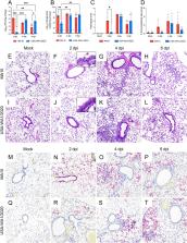
          Several models were developed to study the pathogenicity of severe acute respiratory syndrome coronavirus 2 (SARS-CoV-2) as well as the in vivo efficacy of vaccines and therapeutics. Since wild-type mice are naturally resistant to infection by ancestral SARS-CoV-2 strains, several transgenic mouse models expressing human angiotensin-converting enzyme 2 (hACE2) were developed. An alternative approach has been to develop mouse-adapted SARS-CoV-2 strains. Here, we compared the clinical progression, viral replication kinetics and dissemination, pulmonary tropism, and host innate immune response dynamics between the mouse-adapted MA10 strain and its parental strain (USA-WA1/2020) following intranasal inoculation of K18-hACE2 mice, a widely used model. Compared to its parental counterpart, the MA10 strain induced earlier clinical decline with significantly higher viral replication and earlier neurodissemination. Importantly, the MA10 strain also showed a wider tropism, with infection of bronchiolar epithelia. While both SARS-CoV-2 strains induced comparable pulmonary cytokine/chemokine responses, many proinflammatory and monocyte-recruitment chemokines, such as interleukin-6 (IL-6), tumor necrosis factor alpha (TNF-α), IP-10/CXCL10, and MCP-1/CCL2, showed an earlier peak in MA10-infected mice. Furthermore, both strains induced a similar downregulation of murine Ace2, with only a transient downregulation of Tmprss2 and no alterations in hACE2 expression. Overall, these data demonstrate that in K18-hACE2 mice, the MA10 strain has a pulmonary tropism that more closely resembles SARS-CoV-2 tropism in humans (airways and pneumocytes) than its parental strain. Its rapid replication and neurodissemination and early host pulmonary responses can have a significant impact on the clinical outcomes of infection and are, therefore, critical features to consider for study designs using these strains and mouse model.

          IMPORTANCE The COVID-19 pandemic, caused by SARS-CoV-2, is still significantly impacting health care systems around the globe. Refined animal models are needed to study SARS-CoV-2 pathogenicity as well as efficacy of vaccines and therapeutics. In line with this, thorough evaluation of animal models and virus strains/variants are paramount for standardization and meaningful comparisons. Here, we demonstrated differences in replication dynamics between the Wuhan-like USA-WA1/2020 strain and the derivative mouse-adapted MA10 strain in K18-hACE2 mice. The MA10 strain showed accelerated viral replication and neurodissemination, differential pulmonary tropism, and earlier pulmonary innate immune responses. The observed differences allow us to better refine experimental designs when considering the use of the MA10 strain in the widely utilized K18-hACE2 murine model.

          Related collections

          Most cited references82

          • Record: found
          • Abstract: found
          • Article: found
          Is Open Access

          A pneumonia outbreak associated with a new coronavirus of probable bat origin

          Since the outbreak of severe acute respiratory syndrome (SARS) 18 years ago, a large number of SARS-related coronaviruses (SARSr-CoVs) have been discovered in their natural reservoir host, bats 1–4 . Previous studies have shown that some bat SARSr-CoVs have the potential to infect humans 5–7 . Here we report the identification and characterization of a new coronavirus (2019-nCoV), which caused an epidemic of acute respiratory syndrome in humans in Wuhan, China. The epidemic, which started on 12 December 2019, had caused 2,794 laboratory-confirmed infections including 80 deaths by 26 January 2020. Full-length genome sequences were obtained from five patients at an early stage of the outbreak. The sequences are almost identical and share 79.6% sequence identity to SARS-CoV. Furthermore, we show that 2019-nCoV is 96% identical at the whole-genome level to a bat coronavirus. Pairwise protein sequence analysis of seven conserved non-structural proteins domains show that this virus belongs to the species of SARSr-CoV. In addition, 2019-nCoV virus isolated from the bronchoalveolar lavage fluid of a critically ill patient could be neutralized by sera from several patients. Notably, we confirmed that 2019-nCoV uses the same cell entry receptor—angiotensin converting enzyme II (ACE2)—as SARS-CoV.
            Bookmark
            • Record: found
            • Abstract: found
            • Article: not found

            SARS-CoV-2 Cell Entry Depends on ACE2 and TMPRSS2 and Is Blocked by a Clinically Proven Protease Inhibitor

            Summary The recent emergence of the novel, pathogenic SARS-coronavirus 2 (SARS-CoV-2) in China and its rapid national and international spread pose a global health emergency. Cell entry of coronaviruses depends on binding of the viral spike (S) proteins to cellular receptors and on S protein priming by host cell proteases. Unravelling which cellular factors are used by SARS-CoV-2 for entry might provide insights into viral transmission and reveal therapeutic targets. Here, we demonstrate that SARS-CoV-2 uses the SARS-CoV receptor ACE2 for entry and the serine protease TMPRSS2 for S protein priming. A TMPRSS2 inhibitor approved for clinical use blocked entry and might constitute a treatment option. Finally, we show that the sera from convalescent SARS patients cross-neutralized SARS-2-S-driven entry. Our results reveal important commonalities between SARS-CoV-2 and SARS-CoV infection and identify a potential target for antiviral intervention.
              Bookmark
              • Record: found
              • Abstract: found
              • Article: found
              Is Open Access

              A new coronavirus associated with human respiratory disease in China

              Emerging infectious diseases, such as severe acute respiratory syndrome (SARS) and Zika virus disease, present a major threat to public health 1–3 . Despite intense research efforts, how, when and where new diseases appear are still a source of considerable uncertainty. A severe respiratory disease was recently reported in Wuhan, Hubei province, China. As of 25 January 2020, at least 1,975 cases had been reported since the first patient was hospitalized on 12 December 2019. Epidemiological investigations have suggested that the outbreak was associated with a seafood market in Wuhan. Here we study a single patient who was a worker at the market and who was admitted to the Central Hospital of Wuhan on 26 December 2019 while experiencing a severe respiratory syndrome that included fever, dizziness and a cough. Metagenomic RNA sequencing 4 of a sample of bronchoalveolar lavage fluid from the patient identified a new RNA virus strain from the family Coronaviridae, which is designated here ‘WH-Human 1’ coronavirus (and has also been referred to as ‘2019-nCoV’). Phylogenetic analysis of the complete viral genome (29,903 nucleotides) revealed that the virus was most closely related (89.1% nucleotide similarity) to a group of SARS-like coronaviruses (genus Betacoronavirus, subgenus Sarbecovirus) that had previously been found in bats in China 5 . This outbreak highlights the ongoing ability of viral spill-over from animals to cause severe disease in humans.
                Bookmark

                Author and article information

                Contributors
                Role: Editor
                Journal
                mSphere
                mSphere
                msphere
                mSphere
                American Society for Microbiology (1752 N St., N.W., Washington, DC )
                2379-5042
                2 February 2023
                Jan-Feb 2023
                2 February 2023
                : 8
                : 1
                : e00558-22
                Affiliations
                [a ] Department of Pathobiological Sciences, School of Veterinary Medicine, Louisiana State University, Baton Rouge, Louisiana, USA
                [b ] Louisiana Animal Disease Diagnostic Laboratory, School of Veterinary Medicine, Louisiana State University, Baton Rouge, Louisiana, USA
                [c ] National Emerging Infectious Diseases Laboratories (NEIDL), Boston University, Boston, Massachusetts, USA
                [d ] Department of Pathology and Laboratory Medicine, Boston University Chobanian and Avedisian School of Medicine, Boston, Massachusetts, USA
                [e ] Department of Diagnostic Medicine and Pathobiology, College of Veterinary Medicine, Kansas State University, Manhattan, Kansas, USA
                University of Michigan—Ann Arbor
                Author notes

                The authors declare no conflict of interest.

                Author information
                https://orcid.org/0000-0001-7402-5989
                https://orcid.org/0000-0003-0609-678X
                https://orcid.org/0000-0003-3873-9188
                https://orcid.org/0000-0001-7308-5672
                https://orcid.org/0000-0003-3864-5915
                Article
                00558-22 msphere.00558-22
                10.1128/msphere.00558-22
                9942576
                36728430
                dc7b6b86-0ab5-4f39-8844-08cadf1eafaf
                Copyright © 2023 Thieulent et al.

                This is an open-access article distributed under the terms of the Creative Commons Attribution 4.0 International license.

                History
                : 7 November 2022
                : 3 January 2023
                Page count
                supplementary-material: 5, Figures: 9, Tables: 2, Equations: 0, References: 82, Pages: 23, Words: 14673
                Funding
                Funded by: HHS | National Institutes of Health (NIH), FundRef https://doi.org/10.13039/100000002;
                Award ID: P20GM130555
                Award Recipient :
                Funded by: HHS | National Institutes of Health (NIH), FundRef https://doi.org/10.13039/100000002;
                Award ID: S10-OD030269
                Award Recipient :
                Funded by: LSU | School of Veterinary Medicine, Louisiana State University (SVM, LSU), FundRef https://doi.org/10.13039/100012429;
                Award ID: PG009641
                Award Recipient :
                Funded by: LSU | School of Veterinary Medicine, Louisiana State University (SVM, LSU), FundRef https://doi.org/10.13039/100012429;
                Award ID: PG008671
                Award Recipient :
                Funded by: LSU | School of Veterinary Medicine, Louisiana State University (SVM, LSU), FundRef https://doi.org/10.13039/100012429;
                Award ID: PG008671
                Award Recipient :
                Funded by: U.S. Department of Agriculture (USDA), FundRef https://doi.org/10.13039/100000199;
                Award ID: 2019-67016-29102
                Award Recipient :
                Categories
                Research Article
                virology, Virology
                Custom metadata
                January/February 2023

                ace2-expressing mice,covid-19,k18-hace2 mice,ma10,mouse-adapted virus,respiratory infection,sars-cov-2,wa1,mace2,mouse model,viral replication

                Comments

                Comment on this article