10
views
0
recommends
+1 Recommend
0 collections
    0
    shares
      • Record: found
      • Abstract: found
      • Article: found
      Is Open Access

      Performance of an Automated Fluorescence Antinuclear Antibody Image Analyzer

      research-article

      Read this article at

      Bookmark
          There is no author summary for this article yet. Authors can add summaries to their articles on ScienceOpen to make them more accessible to a non-specialist audience.

          Abstract

          Background

          The gold standard for antinuclear antibody (ANA) screening is the indirect immunofluorescence (IIF) assay with human epithelial cells (HEp-2). However, a number of substantial disadvantages of manual IIF assays have highlighted the need for the automation and standardization of fluorescent ANA (FANA) testing. We evaluated the performance of EUROPattern Suite (Euroimmun AG, Germany), an automated FANA image analyzer, with regard to ANA detection and pattern recognition compared with conventional manual interpretation using the fluorescence microscopic IIF assay.

          Methods

          A total of 104 samples including 70 ANA-positive sera and 34 ANA-negative sera collected from September to October 2015 were included. The sensitivity, specificity, and pattern recognition function were evaluated to determine the performance of EUROPattern Suite compared with the manual IIF assay results.

          Results

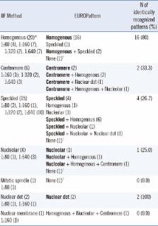
          The sensitivity and specificity of EUROPattern Suite for ANA detection were 94.3% and 94.1%, respectively. The concordance rate between the two methods was 94.2%. For pattern recognition, 45.7% of the samples were assigned identical ANA patterns including simple and mixed. When major pattern matching was considered, 83.7% (41/49) and 95.2% (20/21) of the samples with simple and mixed patterns, respectively, showed concordant results between the two methods.

          Conclusions

          EUROPattern Suite, an automated FANA image analyzer, provides a viable option for distinguishing between positive and negative results, although the ability to assign specific patterns is insufficient to replace manual microscopic interpretation. This automated system may increase efficiency in laboratories, in which a large number of samples need to be processed.

          Related collections

          Most cited references16

          • Record: found
          • Abstract: found
          • Article: not found

          International consensus on ANA patterns (ICAP): the bumpy road towards a consensus on reporting ANA results

          The International Consensus on ANA Patterns (ICAP) was initiated as a workshop aiming to thoroughly discuss and achieve consensus regarding the morphological patterns observed in the indirect immunofluorescence assay on HEp-2 cells. One of the topics discussed at the second ICAP workshop, and addressed in this paper, was the harmonization of reporting ANA test results. This discussion centered on the issue if cytoplasmic and mitotic patterns should be reported as positive or negative. This report outlines the issues that impact on two major different reporting methods. Although it was appreciated by all participants that cytoplasmic and mitotic patterns are clinically relevant, implications for existing diagnostic/classification criteria for ANA-associated diseases in particular hampered a final consensus on this topic. Evidently, a more concerted action of all relevant stakeholders is required. Future ICAP workshops may help to facilitate this action.
            Bookmark
            • Record: found
            • Abstract: found
            • Article: not found

            Importance of the dense fine speckled pattern on HEp-2 cells and anti-DFS70 antibodies for the diagnosis of systemic autoimmune diseases.

            The presence of anti-nuclear antibodies (ANA) is a hallmark of systemic autoimmune rheumatic diseases (SARD). The indirect immunofluorescence (IIF) assay on HEp-2 cells is a commonly used test for the detection of ANA and was recently recommended as the screening test of choice by a task force of the American College of Rheumatology. However, up to 20% of serum samples from healthy individuals (HI) have been reported to have a positive ANA test, the majority of which are directed to the dense fine speckles 70 (DFS70) antigen. Even more important, the DFS IIF pattern has been reported in 33% of ANA positive HI, but not in ANA positive SARD sera. Since the intended use of the ANA HEp-2 test is to aid in the diagnosis of SARD, the reporting of anti-DFS70 antibodies and their associated pattern (DFS) as a positive test, significantly reduces the specificity and the positive likelihood of the ANA test. This has significant implications for diagnostic algorithms involving the detection of ANA. We summarize the current knowledge of anti-DFS70 antibodies and their impact on ANA testing. We also suggest a test algorithm which considers the DFS pattern and the presence of anti-DFS70 antibodies. In addition, we describe a novel method based on immunoadsorption of anti-DFS70 antibodies, which increases the specificity of the ANA HEp-2 test for SARD and which has the potential to overcome a significant limitation of the ANA HEp-2 assay. Copyright © 2011 Elsevier B.V. All rights reserved.
              Bookmark
              • Record: found
              • Abstract: found
              • Article: not found

              Automated antinuclear immunofluorescence antibody screening: a comparative study of six computer-aided diagnostic systems.

              Indirect immunofluorescence (IIF) plays an important role in immunological assays for detecting and measuring autoantibodies. However, the method is burdened by some unfavorable features: the need for expert morphologists, the subjectivity of interpretation, and a low degree of standardization and automation. Following the recent statement by the American College of Rheumatology that the IIF technique should be considered as the standard screening method for the detection of anti-nuclear antibodies (ANA), the biomedical industry has developed technological solutions which might significantly improve automation of the procedure, not only in the preparation of substrates and slides, but also in microscope reading. We collected 104 ANA-positive sera from patients with a confirmed clinical diagnosis of autoimmune disease and 40 ANA-negative sera from healthy blood donors. One aliquot of each serum, without information about pattern and titer, was sent to six laboratories of our group, where the sera were tested with the IIF manual method provided by each of the six manufacturers of automatic systems. Assignment of result (pos/neg), of pattern and titer was made by consensus at a meeting attended by all members of the research team. Result was assigned if consensus for pos/neg was reached by at least four of six certifiers, while for the pattern and for the titer, the value observed with higher frequency (mode) was adopted. Seventeen ANA-positive sera and six ANA-negative sera were excluded. Therefore, the study with the following automatic instrumentation was conducted on 92 ANA-positive sera and on 34 ANA-negative sera: Aklides, EUROPattern, G-Sight (I-Sight-IFA), Helios, Image Navigator, and Nova View. Analytical imprecision was measured in five aliquots of the same serum, randomly added to the sample series. Overall sensitivity of the six automated systems was 96.7% and overall specificity was 89.2%. Most false negatives were recorded for cytoplasmic patterns, whereas among nuclear patterns those with a low level of fluorescence (i.e., multiple nuclear dots, midbody, nuclear rim) were sometimes missed. The intensity values of the light signal of various instruments showed a good correlation with the titer obtained by manual reading (Spearman's rho between 0.672 and 0.839; P<0.0001 for all the systems). Imprecision ranged from 1.99% to 25.2% and, for all the systems, it was lower than that obtained by the manual IIF test (39.1%). The accuracy of pattern recognition, which is for now restricted to the most typical patterns (homogeneous, speckled, nucleolar, centromere, multiple nuclear dots and cytoplasmic) was limited, ranging from 52% to 79%. This study, which is the first to compare the diagnostic accuracy of six systems for automated ANA-IIF reading on the same series of sera, showed that all systems are able to perform very well the task for which they were created. Indeed, cumulative automatic discrimination between positive and negative samples had 95% accuracy. All the manufacturers are actively continuing the development of new and more sophisticated software for a better definition in automatic recognition of patterns and light signal conversion in end-point titer. In the future, this may avert the need for serum dilution for titration, which will be a great advantage in economic terms and time-saving. Copyright © 2013 Elsevier B.V. All rights reserved.
                Bookmark

                Author and article information

                Journal
                Ann Lab Med
                Ann Lab Med
                ALM
                Annals of Laboratory Medicine
                The Korean Society for Laboratory Medicine
                2234-3806
                2234-3814
                May 2017
                17 February 2017
                : 37
                : 3
                : 240-247
                Affiliations
                [1 ]Department of Laboratory Medicine & Genetics, Samsung Medical Center, Sungkyunkwan University School of Medicine, Seoul, Korea.
                [2 ]Department of Internal Medicine, Samsung Medical Center, Sungkyunkwan University School of Medicine, Seoul, Korea.
                Author notes
                Corresponding author: Eun-Suk Kang. Department of Laboratory Medicine & Genetics, Samsung Medical Center, Sungkyunkwan University School of Medicine, 81 Irwon-ro, Gangnam-gu, Seoul 06351, Korea. Tel: +82-2-3410-2703, Fax: +82-2-3410-2719, eskang@ 123456skku.edu
                Article
                10.3343/alm.2017.37.3.240
                5339096
                28224770
                dc1d411b-e95c-4d08-ac12-9ee2c9df176b
                © The Korean Society for Laboratory Medicine

                This is an Open Access article distributed under the terms of the Creative Commons Attribution Non-Commercial License ( http://creativecommons.org/licenses/by-nc/4.0/) which permits unrestricted non-commercial use, distribution, and reproduction in any medium, provided the original work is properly cited.

                History
                : 01 August 2016
                : 08 November 2016
                : 26 January 2017
                Categories
                Original Article
                Diagnostic Immunology

                Clinical chemistry
                antinuclear antibody,indirect immunofluorescence assay,automated image analyzer,pattern recognition

                Comments

                Comment on this article