5
views
0
recommends
+1 Recommend
0 collections
    0
    shares
      • Record: found
      • Abstract: found
      • Article: found
      Is Open Access

      CFTR Therapeutics Normalize Cerebral Perfusion Deficits in Mouse Models of Heart Failure and Subarachnoid Hemorrhage

      research-article
      , PhD a , b , , , MSc a , b , , , PhD c , , MSc a , b , , PhD a , b , , PhD a , b , , PhD a , , PhD c , , MSc c , , BSc a , , MD, PhD d , , MD a , b , , PhD e , , PhD f , , BSc g , h , i , , MD h , j , , MD, PhD g , h , , MD a , d , k , l , , PhD, DVM e , m , , PhD a , k , , PhD a , c , , , MD, PhD a , b , k , ,
      JACC: Basic to Translational Science
      Elsevier
      cognitive impairment, corrector compounds, cystic fibrosis transmembrane conductance regulator (CFTR), myogenic vasoconstriction, sphingosine-1-phosphate, tumor necrosis factor, vascular smooth muscle cells, CBF, cerebral blood flow, CFTR, cystic fibrosis transmembrane conductance regulator, HF, heart failure, MAP, mean arterial pressure, MOPS, 3-morpholinopropanesulfonic acid, MRI, magnetic resonance imaging, NIH, National Institutes of Health, PCA, posterior cerebral artery, SAH, subarachnoid hemorrhage, S1P, sphingosine-1-phosphate, TNF, tumor necrosis factor, TPR, total peripheral resistance

      Read this article at

      Bookmark
          There is no author summary for this article yet. Authors can add summaries to their articles on ScienceOpen to make them more accessible to a non-specialist audience.

          Visual Abstract

          Highlights

          • The cystic fibrosis transmembrane conductance regulator (CFTR) is a significant modulator of cerebrovascular reactivity; the loss of CFTR function enhances myogenic vasoconstriction.

          • Heart failure and subarachnoid hemorrhage downregulate cerebrovascular CFTR protein expression; this leads to enhanced cerebral artery vasoconstriction, reduced cerebral perfusion, neuronal injury, and ultimately, neurologic deficits.

          • CFTR therapeutics that maintain CFTR expression normalize the perfusion deficits, reduce neuronal injury, and improve neurologic function in these pathological settings.

          Summary

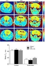
          Heart failure (HF) and subarachnoid hemorrhage (SAH) chronically reduce cerebral perfusion, which negatively affects clinical outcome. This work demonstrates a strong relationship between cerebral artery cystic fibrosis transmembrane conductance regulator (CFTR) expression and altered cerebrovascular reactivity in HF and SAH. In HF and SAH, CFTR corrector compounds (C18 or lumacaftor) normalize pathological alterations in cerebral artery CFTR expression, vascular reactivity, and cerebral perfusion, without affecting systemic hemodynamic parameters. This normalization correlates with reduced neuronal injury. Therefore, CFTR therapeutics have emerged as valuable clinical tools to manage cerebrovascular dysfunction, impaired cerebral perfusion, and neuronal injury.

          Related collections

          Most cited references25

          • Record: found
          • Abstract: found
          • Article: not found

          Novel Object Recognition Test for the Investigation of Learning and Memory in Mice

          The object recognition test (ORT) is a commonly used behavioral assay for the investigation of various aspects of learning and memory in mice. The ORT is fairly simple and can be completed over 3 days: habituation day, training day, and testing day. During training, the mouse is allowed to explore 2 identical objects. On test day, one of the training objects is replaced with a novel object. Because mice have an innate preference for novelty, if the mouse recognizes the familiar object, it will spend most of its time at the novel object. Due to this innate preference, there is no need for positive or negative reinforcement or long training schedules. Additionally, the ORT can also be modified for numerous applications. The retention interval can be shortened to examine short-term memory, or lengthened to probe long-term memory. Pharmacological intervention can be used at various times prior to training, after training, or prior to recall to investigate different phases of learning ( i.e. , acquisition, early or late consolidation, or recall). Overall, the ORT is a relatively low-stress, efficient test for memory in mice, and is appropriate for the detection of neuropsychological changes following pharmacological, biological, or genetic manipulations.
            Bookmark
            • Record: found
            • Abstract: found
            • Article: not found

            Cerebral autoregulation.

            Autoregulation of blood flow denotes the intrinsic ability of an organ or a vascular bed to maintain a constant perfusion in the face of blood pressure changes. Alternatively, autoregulation can be defined in terms of vascular resistance changes or simply arteriolar caliber changes as blood pressure or perfusion pressure varies. While known in almost any vascular bed, autoregulation and its disturbance by disease has attracted particular attention in the cerebrovascular field. The basic mechanism of autoregulation of cerebral blood flow (CBF) is controversial. Most likely, the autoregulatory vessel caliber changes are mediated by an interplay between myogenic and metabolic mechanisms. Influence of perivascular nerves and most recently the vascular endothelium has also been the subject of intense investigation. CBF autoregulation typically operates between mean blood pressures of the order of 60 and 150 mm Hg. These limits are not entirely fixed but can be modulated by sympathetic nervous activity, the vascular renin-angiotensin system, and any factor (notably changes in arterial carbon dioxide tension) that decreases or increases CBF. Disease states of the brain may impair or abolish CBF autoregulation. Thus, autoregulation is lost in severe head injury or acute ischemic stroke, leaving surviving brain tissue unprotected against the potentially harmful effect of blood pressure changes. Likewise, autoregulation may be lost in the surroundings of a space-occupying brain lesion, be it a tumor or a hematoma. In many such disease states, autoregulation may be regained by hyperventilatory hypocapnia. Autoregulation may also be impaired in neonatal brain asphyxia and infections of the central nervous system, but appears to be intact in spreading depression and migraine, despite impairment of chemical and metabolic control of CBF. In chronic hypertension, the limits of autoregulation are shifted toward high blood pressure. Acute hypertensive encephalopathy, on the other hand, is thought to be due to autoregulatory failure at very high pressure. In long-term diabetes mellitus there may be chronic impairment of CBF autoregulation, probably due to diabetic microangiopathy.
              Bookmark
              • Record: found
              • Abstract: found
              • Article: not found

              Chronic stress alters dendritic morphology in rat medial prefrontal cortex.

              Chronic stress produces deficits in cognition accompanied by alterations in neural chemistry and morphology. Medial prefrontal cortex is a target for glucocorticoids involved in the stress response. We have previously demonstrated that 3 weeks of daily corticosterone injections result in dendritic reorganization in pyramidal neurons in layer II-III of medial prefrontal cortex. To determine if similar morphological changes occur in response to chronic stress, we assessed the effects of daily restraint stress on dendritic morphology in medial prefrontal cortex. Male rats were exposed to either 3 h of restraint stress daily for 3 weeks or left unhandled except for weighing during this period. On the last day of restraint, animals were overdosed and brains were stained using a Golgi-Cox procedure. Pyramidal neurons in lamina II-III of medial prefrontal cortex were drawn in three dimensions, and the morphology of apical and basilar arbors was quantified. Sholl analyses demonstrated a significant alteration of apical dendrites in stressed animals: overall, the number and length of apical dendritic branches was reduced by 18 and 32%, respectively. The reduction in apical dendritic arbor was restricted to distal and higher-order branches, and may reflect atrophy of terminal branches: terminal branch number and length were reduced by 19 and 35%. On the other hand, basilar dendrites were not affected. This pattern of dendritic reorganization is similar to that seen after daily corticosterone injections. This reorganization likely reflects functional changes in prefrontal cortex and may contribute to stress-induced changes in cognition. Copyright 2004 Wiley Periodicals, Inc.
                Bookmark

                Author and article information

                Contributors
                Journal
                JACC Basic Transl Sci
                JACC Basic Transl Sci
                JACC: Basic to Translational Science
                Elsevier
                2452-302X
                27 November 2019
                December 2019
                27 November 2019
                : 4
                : 8
                : 940-958
                Affiliations
                [a ]Department of Physiology, University of Toronto, Toronto, Ontario, Canada
                [b ]Toronto Centre for Microvascular Medicine at The Ted Rogers Centre for Heart Research Translational Biology and Engineering Program, University of Toronto, Ontario, Canada
                [c ]Wallenberg Center for Molecular Medicine and Department of Experimental Medical Science, Lund University, Lund, Sweden
                [d ]Division of Cell & Molecular Biology, Toronto General Hospital Research Institute, Toronto, Ontario, Canada
                [e ]Division of Cardiology, University Health Network, Toronto, Ontario, Canada
                [f ]STTARR Innovation Centre, Department of Radiation Oncology, Princess Margaret Hospital, Toronto, Ontario, Canada
                [g ]Labatt Family Centre of Excellence in Brain Injury and Trauma Research, Keenan Research Centre for Biomedical Research and Li Ka Shing Knowledge Institute, St. Michael’s Hospital, Toronto, Ontario, Canada
                [h ]Division of Neurosurgery, St. Michael’s Hospital, and Department of Surgery, University of Toronto, Toronto, Ontario, Canada
                [i ]Sunnybrook Research Institute, Physical Sciences Platform and Department of Medical Biophysics, University of Toronto, Toronto, Ontario, Canada
                [j ]Institute of Health Biosciences, Department of Neurosurgery, University of Tokushima Graduate School, Tokushima, Japan
                [k ]Heart & Stroke/Richard Lewar Centre of Excellence for Cardiovascular Research, University of Toronto, Toronto, Ontario, Canada
                [l ]Department of Medicine, University of Toronto, Toronto, Ontario, Canada
                [m ]Department of Biology, York University, Toronto, Ontario, Canada
                Author notes
                [] Address for correspondence: Dr. Steffen-Sebastian Bolz, Toronto Centre for Microvascular Medicine at TBEP, University of Toronto, 661 University Avenue, 14th Floor, Toronto, Ontario, Canada, M5G 1M1. sts.bolz@ 123456utoronto.ca
                [∗]

                Dr. Lidington and Ms. Fares contributed equally to this work and are joint first authors.

                [†]

                Drs. Meissner and Bolz contributed equally to this work and are joint senior authors.

                Article
                S2452-302X(19)30194-9
                10.1016/j.jacbts.2019.07.004
                6939007
                31909302
                db61c3e0-c028-4bb6-92cc-3cd8972fbfce
                © 2019 The Authors

                This is an open access article under the CC BY-NC-ND license (http://creativecommons.org/licenses/by-nc-nd/4.0/).

                History
                : 29 April 2019
                : 15 July 2019
                : 16 July 2019
                Categories
                Editorial Comment

                cognitive impairment,corrector compounds,cystic fibrosis transmembrane conductance regulator (cftr),myogenic vasoconstriction,sphingosine-1-phosphate,tumor necrosis factor,vascular smooth muscle cells,cbf, cerebral blood flow,cftr, cystic fibrosis transmembrane conductance regulator,hf, heart failure,map, mean arterial pressure,mops, 3-morpholinopropanesulfonic acid,mri, magnetic resonance imaging,nih, national institutes of health,pca, posterior cerebral artery,sah, subarachnoid hemorrhage,s1p, sphingosine-1-phosphate,tnf, tumor necrosis factor,tpr, total peripheral resistance

                Comments

                Comment on this article