14
views
0
recommends
+1 Recommend
0 collections
    0
    shares
      • Record: found
      • Abstract: found
      • Article: found
      Is Open Access

      Sinus mucosal healing pattern according to pterygomaxillary disjunction type after Le Fort I osteotomy

      research-article

      Read this article at

      Bookmark
          There is no author summary for this article yet. Authors can add summaries to their articles on ScienceOpen to make them more accessible to a non-specialist audience.

          Abstract

          Objectives

          During Le Fort I osteotomy, the separation of the pterygomaxillary junction (PMJ) is a difficult procedure for most surgeons because it is invisible. In this process, damage to the posterior structures constituting the sinus or those adjacent to it, including the maxillary sinus posterior wall and pterygoid plate, may occur. We would like to investigate the effects of this on the inside of the maxillary sinus after surgery and whether there are complications.

          Materials and Methods

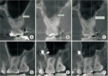
          One-hundred patients who underwent Le Fort I osteotomy from 2013 to 2020 using cone-beam computed tomography images were classified into two groups (clean-cut type and fractured type) according to the PMJ cutting pattern. In addition, the mucosal thickness in the maxillary sinus was divided into preoperative, postoperative three months, one year, and the change over the course of surgery was evaluated retrospectively.

          Results

          Of the total 100 cases, the clean-cut type numbered 28 cases and the fractured type totaled 72 cases. Among the fracture types, part of the sinus wall and the pterygoid plate were broken in 69 cases, and the maxillary sinus posterior wall was detached in three cases. There was no statistically significant difference in sinus mucosal thickening between the clean-cut type and fractured type of the PMJ, three months and one year after surgery between the two groups. However, there was a significant difference in sinus mucosal thickness at postoperative one year in the case where a partial detachment of the maxillary sinus posterior wall occurred compared to not.

          Conclusion

          Even if there is some damage to the structures behind the PMJ, it may not be reasonable to spend some time on the PMJ separation process considering the overall postoperative complications, if there is no significant difference inside the sinus, or increased probability of postoperative complications.

          Related collections

          Most cited references7

          • Record: found
          • Abstract: found
          • Article: not found

          Association between periapical lesions and maxillary sinus mucosal thickening: a retrospective cone-beam computed tomographic study.

          Odontogenic infections are a common cause of maxillary sinusitis. This study aimed to evaluate the relationship between teeth with periapical lesions or periodontal disease and sinus mucosal thickening using cone-beam computed tomography (CBCT) imaging.
            Bookmark
            • Record: found
            • Abstract: found
            • Article: not found

            Dimensional changes of maxillary sinuses and pharyngeal airway in Class III patients undergoing bimaxillary orthognathic surgery.

            To evaluate the pharyngeal airway and maxillary sinus volume changes after mandibular setback surgery combined with maxillary advancement and/or impaction surgery.
              Bookmark
              • Record: found
              • Abstract: found
              • Article: not found

              Preoperative and Postoperative CT Scan Assessment of Pterygomaxillary Junction in Patients Undergoing Le Fort I Osteotomy: Comparison of Pterygomaxillary Dysjunction Technique and Trimble Technique-A Pilot Study.

              To determine the rate of complications and occurrence of pterygoid plate fractures comparing two techniques of Le Fort I osteotomy i.e., Classic Pterygomaxillary Dysjunction technique and Trimble technique and to know whether the dimensions of pterygomaxillary junction [determined preoperatively by computed tomography (CT) scan] have any influence on pterygomaxillary separation achieved during surgery.
                Bookmark

                Author and article information

                Journal
                J Korean Assoc Oral Maxillofac Surg
                J Korean Assoc Oral Maxillofac Surg
                Journal of the Korean Association of Oral and Maxillofacial Surgeons
                The Korean Association of Oral and Maxillofacial Surgeons
                2234-7550
                2234-5930
                31 October 2022
                31 October 2022
                31 October 2022
                : 48
                : 5
                : 292-296
                Affiliations
                [1 ]Department of Oral and Maxillofacial Surgery, Kyung Hee University Dental Hospital, Seoul, Korea
                [2 ]Department of Oral and Maxillofacial Surgery, School of Dentistry, Kyung Hee University, Seoul, Korea
                Author notes
                Joo-Young Ohe, Department of Oral and Maxillofacial Surgery, Kyung Hee University Dental Hospital, 26 Kyungheedae-ro, Dongdaemun-gu, Seoul 02447, Korea, TEL: +82-2-958-9360, E-mail: ojyoung81@ 123456naver.com , ORCID: https://orcid.org/0000-0002-4411-7871
                Author information
                https://orcid.org/0000-0001-9063-9761
                https://orcid.org/0000-0001-6750-9643
                https://orcid.org/0000-0002-9145-8062
                https://orcid.org/0000-0003-1938-1846
                https://orcid.org/0000-0002-2870-2747
                https://orcid.org/0000-0002-4411-7871
                Article
                jkaoms-48-5-292
                10.5125/jkaoms.2022.48.5.292
                9639255
                36316187
                d90bf410-08ac-4dd4-a681-68d19acf7287
                Copyright © 2022 The Korean Association of Oral and Maxillofacial Surgeons. All rights reserved.

                This is an open-access article distributed under the terms of the Creative Commons Attribution Non-Commercial License ( http://creativecommons.org/licenses/by-nc/4.0), which permits unrestricted non-commercial use, distribution, and reproduction in any medium, provided the original work is properly cited.

                History
                : 19 July 2022
                : 11 August 2022
                : 17 August 2022
                Categories
                Original Article

                maxillary osteotomy,pterygomaxillary junction,maxillary sinus,mucous membrane

                Comments

                Comment on this article