57
views
0
recommends
+1 Recommend
0 collections
    0
    shares
      • Record: found
      • Abstract: found
      • Article: found
      Is Open Access

      Diffuse Decreased Gray Matter in Patients with Idiopathic Craniocervical Dystonia: A Voxel-Based Morphometry Study

      research-article

      Read this article at

      Bookmark
          There is no author summary for this article yet. Authors can add summaries to their articles on ScienceOpen to make them more accessible to a non-specialist audience.

          Abstract

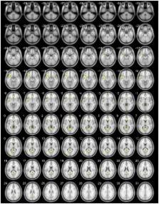
          Background: Recent studies have addressed the role of structures other than the basal ganglia in the pathophysiology of craniocervical dystonia (CCD). Neuroimaging studies have attempted to identify structural abnormalities in CCD but a clear pattern of alteration has not been established. We performed whole-brain evaluation using voxel-based morphometry (VBM) to identify patterns of gray matter (GM) changes in CCD.

          Methods: We compared 27 patients with CCD matched in age and gender to 54 healthy controls. VBM was used to compare GM volumes. We created a two-sample t-test corrected for subjects’ age, and we tested with a level of significance of p < 0.001 and false discovery rate (FDR) correction ( p < 0.05).

          Results: Voxel-based morphometry demonstrated significant reductions of GM using p < 0.001 in the cerebellar vermis IV/V, bilaterally in the superior frontal gyrus, precuneus, anterior cingulate and paracingulate, insular cortex, lingual gyrus, and calcarine fissure; in the left hemisphere in the supplementary motor area, inferior frontal gyrus, inferior parietal gyrus, temporal pole, supramarginal gyrus, rolandic operculum, hippocampus, middle occipital gyrus, cerebellar lobules IV/V, superior, and middle temporal gyri; in the right hemisphere, the middle cingulate and precentral gyrus. Our study did not report any significant result using the FDR correction. We also detected correlations between GM volume and age, disease duration, duration of botulinum toxin treatment, and the Marsden–Fahn dystonia scale scores.

          Conclusion: We detected large clusters of GM changes chiefly in structures primarily involved in sensorimotor integration, motor planning, visuospatial function, and emotional processing.

          Related collections

          Most cited references35

          • Record: found
          • Abstract: found
          • Article: found
          Is Open Access

          Three Systems of Insular Functional Connectivity Identified with Cluster Analysis

          Despite much research on the function of the insular cortex, few studies have investigated functional subdivisions of the insula in humans. The present study used resting-state functional connectivity magnetic resonance imaging (MRI) to parcellate the human insular lobe based on clustering of functional connectivity patterns. Connectivity maps were computed for each voxel in the insula based on resting-state functional MRI (fMRI) data and segregated using cluster analysis. We identified 3 insular subregions with distinct patterns of connectivity: a posterior region, functionally connected with primary and secondary somatomotor cortices; a dorsal anterior to middle region, connected with dorsal anterior cingulate cortex, along with other regions of a previously described control network; and a ventral anterior region, primarily connected with pregenual anterior cingulate cortex. Applying these regions to a separate task data set, we found that dorsal and ventral anterior insula responded selectively to disgusting images, while posterior insula did not. These results demonstrate that clustering of connectivity patterns can be used to subdivide cerebral cortex into anatomically and functionally meaningful subregions; the insular regions identified here should be useful in future investigations on the function of the insula.
            Bookmark
            • Record: found
            • Abstract: not found
            • Article: not found

            Voxel-Based Morphometry of the Human Brain: Methods and Applications

              Bookmark
              • Record: found
              • Abstract: found
              • Article: not found

              Interactions between frontal cortex and basal ganglia in working memory: a computational model.

              The frontal cortex and the basal ganglia interact via a relatively well understood and elaborate system of interconnections. In the context of motor function, these interconnections can be understood as disinhibiting, or "releasing the brakes," on frontal motor action plans: The basal ganglia detect appropriate contexts for performing motor actions and enable the frontal cortex to execute such actions at the appropriate time. We build on this idea in the domain of working memory through the use of computational neural network models of this circuit. In our model, the frontal cortex exhibits robust active maintenance, whereas the basal ganglia contribute a selective, dynamic gating function that enables frontal memory representations to be rapidly updated in a task-relevant manner. We apply the model to a novel version of the continuous performance task that requires subroutine-like selective working memory updating and compare and contrast our model with other existing models and theories of frontal-cortex-basal-ganglia interactions.
                Bookmark

                Author and article information

                Contributors
                URI : http://frontiersin.org/people/u/179481
                URI : http://frontiersin.org/people/u/201115
                URI : http://frontiersin.org/people/u/201094
                URI : http://frontiersin.org/people/u/201136
                URI : http://frontiersin.org/people/u/117269
                URI : http://frontiersin.org/people/u/187851
                URI : http://frontiersin.org/people/u/201142
                URI : http://frontiersin.org/people/u/201106
                URI : http://frontiersin.org/people/u/201105
                URI : http://frontiersin.org/people/u/66908
                URI : http://frontiersin.org/people/u/55511
                URI : http://frontiersin.org/people/u/8789
                URI : http://frontiersin.org/people/u/72124
                Journal
                Front Neurol
                Front Neurol
                Front. Neurol.
                Frontiers in Neurology
                Frontiers Media S.A.
                1664-2295
                08 January 2015
                2014
                : 5
                : 283
                Affiliations
                [1] 1Neuroimaging Laboratory, University of Campinas , Campinas, Brazil
                [2] 2Department of Neurology, University of Campinas , Campinas, Brazil
                [3] 3Department of Medical Genetics, University of Campinas , Campinas, Brazil
                [4] 4Department of Radiology, University of Campinas , Campinas, Brazil
                Author notes

                Edited by: Antonio Pisani, Università degli Studi di Roma Tor Vergata, Italy

                Reviewed by: Paolo Calabresi, Santa Maria della Misericordia Hospital, Italy; Francesca Morgante, University of Messina, Italy; Giacomo Koch, Fondazione Santa Lucia IRCCS, Italy; Héctor Alberto González-Usigli, Instituto Mexicano del Seguro Social, Mexico

                *Correspondence: Camila C. Piccinin, Neuroimaging Laboratory, School of Medical Sciences, University of Campinas, Cidade Universitária, Campinas, São Paulo 13083-887, Brazil e-mail: camila.piccinin@ 123456gmail.com

                This article was submitted to Movement Disorders, a section of the journal Frontiers in Neurology.

                Article
                10.3389/fneur.2014.00283
                4288053
                25620953
                d8d4bfea-5027-4200-b7da-405e5dc98460
                Copyright © 2015 Piccinin, Piovesana, Santos, Guimarães, De Campos, Rezende, Campos, Torres, Amato-Filho, França, Lopes-Cendes, Cendes and D’Abreu.

                This is an open-access article distributed under the terms of the Creative Commons Attribution License (CC BY). The use, distribution or reproduction in other forums is permitted, provided the original author(s) or licensor are credited and that the original publication in this journal is cited, in accordance with accepted academic practice. No use, distribution or reproduction is permitted which does not comply with these terms.

                History
                : 04 October 2014
                : 14 December 2014
                Page count
                Figures: 5, Tables: 5, Equations: 0, References: 47, Pages: 11, Words: 6575
                Categories
                Neuroscience
                Original Research

                Neurology
                craniocervical dystonia,voxel-based morphometry,gray matter,cervical dystonia,segmental dystonia,neuroimaging

                Comments

                Comment on this article