14
views
0
recommends
+1 Recommend
0 collections
    0
    shares
      • Record: found
      • Abstract: found
      • Article: found
      Is Open Access

      Soman (GD) Rat Model to Mimic Civilian Exposure to Nerve Agent: Mortality, Video-EEG Based Status Epilepticus Severity, Sex Differences, Spontaneously Recurring Seizures, and Brain Pathology

      research-article

      Read this article at

      Bookmark
          There is no author summary for this article yet. Authors can add summaries to their articles on ScienceOpen to make them more accessible to a non-specialist audience.

          Abstract

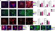
          Modeling a real-world scenario of organophosphate nerve agent (OPNA) exposure is challenging. Military personnel are premedicated with pyridostigmine, which led to the development of OPNA models with pyridostigmine/oxime pretreatment to investigate novel therapeutics for acute and chronic effects. However, civilians are not premedicated with pyridostigmine/oxime. Therefore, experimental models without pyridostigmine were developed by other laboratories though often only in males. Following OPNA exposure, prolonged convulsive seizures (CS) or status epilepticus (SE) are concerning. The duration and severity of CS/SE determine the extent of brain injury in survivors even after treating with medical countermeasures (MCM)/antidotes such as atropine, an oxime, and an anticonvulsant such as diazepam/midazolam. In this study, using a large mixed sex cohort of adult male and female rats, without pretreatment, we demonstrate severe SE lasting for >20 min in 82% of the animals in response to soman (GD,132 μg/kg, s.c.). Atropine sulfate (2 mg/kg, i.m.) and HI-6 (125 mg/kg, i.m.) were administered immediately following soman, and midazolam (3 mg/kg, i.m.) 1 h post-exposure. Immediate MCM treatment is impractical in civilian exposure to civilians, but this approach reduces mortality in experimental models. Interestingly, female rats, irrespective of estrous stages, had an average of 44 min CS (stage ≥ 3), while males had an average of 32 min CS during SE, starting from soman exposure to midazolam treatment. However, in telemetry device implanted groups, there were no significant sex differences in SE severity; males had 40 min and females 43 min of continuous CS until midazolam was administered. No animals died prior to midazolam administration and less than 5% died in the first week after soman intoxication. In telemetered animals, there was a direct correlation between EEG changes and behavioral seizures in real-time. In the long-term, convulsive spontaneously recurring seizures (SRS) were observed in 85% of randomly chosen animals. At 4-months post-soman, the brain histology confirmed reactive gliosis and neurodegeneration. The novel findings of this study are that, in non-telemetered animals, the SE severity following soman intoxication was significantly greater in females compared to males and that the estrous cycle did not influence the response.

          Related collections

          Most cited references97

          • Record: found
          • Abstract: found
          • Article: found
          Is Open Access

          Acetylcholinesterase Inhibitors: Pharmacology and Toxicology

          Acetylcholinesterase is involved in the termination of impulse transmission by rapid hydrolysis of the neurotransmitter acetylcholine in numerous cholinergic pathways in the central and peripheral nervous systems. The enzyme inactivation, induced by various inhibitors, leads to acetylcholine accumulation, hyperstimulation of nicotinic and muscarinic receptors, and disrupted neurotransmission. Hence, acetylcholinesterase inhibitors, interacting with the enzyme as their primary target, are applied as relevant drugs and toxins. This review presents an overview of toxicology and pharmacology of reversible and irreversible acetylcholinesterase inactivating compounds. In the case of reversible inhibitors being commonly applied in neurodegenerative disorders treatment, special attention is paid to currently approved drugs (donepezil, rivastigmine and galantamine) in the pharmacotherapy of Alzheimer’s disease, and toxic carbamates used as pesticides. Subsequently, mechanism of irreversible acetylcholinesterase inhibition induced by organophosphorus compounds (insecticides and nerve agents), and their specific and nonspecific toxic effects are described, as well as irreversible inhibitors having pharmacological implementation. In addition, the pharmacological treatment of intoxication caused by organophosphates is presented, with emphasis on oxime reactivators of the inhibited enzyme activity administering as causal drugs after the poisoning. Besides, organophosphorus and carbamate insecticides can be detoxified in mammals through enzymatic hydrolysis before they reach targets in the nervous system. Carboxylesterases most effectively decompose carbamates, whereas the most successful route of organophosphates detoxification is their degradation by corresponding phosphotriesterases.
            Bookmark
            • Record: found
            • Abstract: found
            • Article: not found

            Reactive microgliosis.

            Damage to the central nervous system (CNS) elicits the activation of both astrocytes and microglia. This review is focused on the principal features that characterize the activation of microglia after CNS injury. It provides a critical discussion of concepts regarding microglial biology that include the relationship between microglia and macrophages, as well as the role of microglia as immunocompetent cells of the CNS. Mechanistic and functional aspects of microgliosis are discussed primarily in the context of microglial neuronal interactions. The controversial issue of whether reactive microgliosis is a beneficial or a harmful process is addressed, and a resolution of this dilemma is offered by suggesting different interpretations of the term 'activated microglia' depending on its usage during in vivo or in vitro experimentation.
              Bookmark
              • Record: found
              • Abstract: found
              • Article: found
              Is Open Access

              An update on reactive astrocytes in chronic pain

              Chronic pain is a critical clinical problem with an increasing prevalence. However, there are limited effective prevention measures and treatments for chronic pain. Astrocytes are the most abundant glial cells in the central nervous system and play important roles in both physiological and pathological conditions. Over the past few decades, a growing body of evidence indicates that astrocytes are involved in the regulation of chronic pain. Recently, reactive astrocytes were further classified into A1 astrocytes and A2 astrocytes according to their functions. After nerve injury, A1 astrocytes can secrete neurotoxins that induce rapid death of neurons and oligodendrocytes, whereas A2 astrocytes promote neuronal survival and tissue repair. These findings can well explain the dual effects of reactive astrocytes in central nervous injury and diseases. In this review, we will summarise the (1) changes in the morphology and function of astrocytes after noxious stimulation and nerve injury, (2) molecular regulators and signalling mechanisms involved in the activation of astrocytes and chronic pain, (3) the role of spinal and cortical astrocyte activation in chronic pain, and (4) the roles of different subtypes of reactive astrocytes (A1 and A2 phenotypes) in nerve injury that is associated with chronic pain. This review provides updated information on the role of astrocytes in the regulation of chronic pain. In particular, we discuss recent findings about A1 and A2 subtypes of reactive astrocytes and make several suggestions for potential therapeutic targets for chronic pain.
                Bookmark

                Author and article information

                Contributors
                Journal
                Front Cell Neurosci
                Front Cell Neurosci
                Front. Cell. Neurosci.
                Frontiers in Cellular Neuroscience
                Frontiers Media S.A.
                1662-5102
                07 February 2022
                2021
                : 15
                : 798247
                Affiliations
                [1] 1Neuroscience Interdepartmental Program, Iowa State University , Ames, IA, United States
                [2] 2Department of Biomedical Sciences, College of Veterinary Medicine, Iowa State University , Ames, IA, United States
                Author notes

                Edited by: Souvarish Sarkar, Brigham and Women’s Hospital and Harvard Medical School, United States

                Reviewed by: Pamela J. Lein, University of California, Davis, United States; Ashish Dhir, University of California, Davis, United States

                *Correspondence: Thimmasettappa Thippeswamy, tswamy@ 123456iastate.edu

                This article was submitted to Cellular Neuropathology, a section of the journal Frontiers in Cellular Neuroscience

                Article
                10.3389/fncel.2021.798247
                8859837
                35197823
                d79db0a8-e75e-4233-b1f1-bfd739791c1e
                Copyright © 2022 Gage, Rao, Samidurai, Putra, Vasanthi, Meyer, Wang and Thippeswamy.

                This is an open-access article distributed under the terms of the Creative Commons Attribution License (CC BY). The use, distribution or reproduction in other forums is permitted, provided the original author(s) and the copyright owner(s) are credited and that the original publication in this journal is cited, in accordance with accepted academic practice. No use, distribution or reproduction is permitted which does not comply with these terms.

                History
                : 19 October 2021
                : 27 December 2021
                Page count
                Figures: 6, Tables: 1, Equations: 0, References: 98, Pages: 15, Words: 11328
                Funding
                Funded by: National Institutes of Health , doi 10.13039/100000002;
                Award ID: 1U01 NS117284
                Award ID: R21 NS110648
                Categories
                Neuroscience
                Original Research

                Neurosciences
                nerve agent,status epilepticus severity,sex as a biological variable,telemetry,mixed-sex cohort,medical countermeasures

                Comments

                Comment on this article