1
views
0
recommends
+1 Recommend
0 collections
    0
    shares
      • Record: found
      • Abstract: found
      • Article: found
      Is Open Access

      Disease-modifying effects of a glial-targeted inducible nitric oxide synthase inhibitor (1400W) in mixed-sex cohorts of a rat soman (GD) model of epilepsy

      research-article

      Read this article at

      Bookmark
          There is no author summary for this article yet. Authors can add summaries to their articles on ScienceOpen to make them more accessible to a non-specialist audience.

          Abstract

          Background

          Acute exposure to seizurogenic organophosphate (OP) nerve agents (OPNA) such as diisopropylfluorophosphate (DFP) or soman (GD), at high concentrations, induce immediate status epilepticus (SE), reactive gliosis, neurodegeneration, and epileptogenesis as a consequence. Medical countermeasures (MCMs—atropine, oximes, benzodiazepines), if administered in < 20 min of OPNA exposure, can control acute symptoms and mortality. However, MCMs alone are inadequate to prevent OPNA-induced brain injury and behavioral dysfunction in survivors. We have previously shown that OPNA exposure-induced SE increases the production of inducible nitric oxide synthase (iNOS) in glial cells in both short- and long- terms. Treating with a water soluble and highly selective iNOS inhibitor, 1400W, for 3 days significantly reduced OPNA-induced brain changes in those animals that had mild–moderate SE in the rat DFP model. However, such mitigating effects and the mechanisms of 1400W are unknown in a highly volatile nerve agent GD exposure.

          Methods

          Mixed-sex cohort of adult Sprague Dawley rats were exposed to GD (132 μg/kg, s.c.) and immediately treated with atropine (2 mg/kg, i.m) and HI-6 (125 mg/kg, i.m.). Severity of seizures were quantified for an hour and treated with midazolam (3 mg/kg, i.m.). An hour post-midazolam, 1400W (20 mg/kg, i.m.) or vehicle was administered daily for 2 weeks. After behavioral testing and EEG acquisition, animals were euthanized at 3.5 months post-GD. Brains were processed for neuroinflammatory and neurodegeneration markers. Serum and CSF were used for nitrooxidative and proinflammatory cytokines assays.

          Results

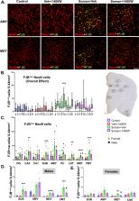
          We demonstrate a significant long-term (3.5 months post-soman) disease-modifying effect of 1400W in animals that had severe SE for > 20 min of continuous convulsive seizures. 1400W significantly reduced GD-induced motor and cognitive dysfunction; nitrooxidative stress (nitrite, ROS; increased GSH: GSSG); proinflammatory cytokines in the serum and some in the cerebrospinal fluid (CSF); epileptiform spikes and spontaneously recurring seizures (SRS) in males; reactive gliosis (GFAP + C3 and IBA1 + CD68-positive glia) as a measure of neuroinflammation, and neurodegeneration (especially parvalbumin-positive neurons) in some brain regions.

          Conclusion

          These findings demonstrate the long-term disease-modifying effects of a glial-targeted iNOS inhibitor, 1400W, in a rat GD model by modulating reactive gliosis, neurodegeneration (parvalbumin-positive neurons), and neuronal hyperexcitability.

          Supplementary Information

          The online version contains supplementary material available at 10.1186/s12974-023-02847-1.

          Related collections

          Most cited references82

          • Record: found
          • Abstract: found
          • Article: not found

          The classical complement cascade mediates CNS synapse elimination.

          During development, the formation of mature neural circuits requires the selective elimination of inappropriate synaptic connections. Here we show that C1q, the initiating protein in the classical complement cascade, is expressed by postnatal neurons in response to immature astrocytes and is localized to synapses throughout the postnatal CNS and retina. Mice deficient in complement protein C1q or the downstream complement protein C3 exhibit large sustained defects in CNS synapse elimination, as shown by the failure of anatomical refinement of retinogeniculate connections and the retention of excess retinal innervation by lateral geniculate neurons. Neuronal C1q is normally downregulated in the adult CNS; however, in a mouse model of glaucoma, C1q becomes upregulated and synaptically relocalized in the adult retina early in the disease. These findings support a model in which unwanted synapses are tagged by complement for elimination and suggest that complement-mediated synapse elimination may become aberrantly reactivated in neurodegenerative disease.
            Bookmark
            • Record: found
            • Abstract: found
            • Article: not found

            PKSolver: An add-in program for pharmacokinetic and pharmacodynamic data analysis in Microsoft Excel.

            This study presents PKSolver, a freely available menu-driven add-in program for Microsoft Excel written in Visual Basic for Applications (VBA), for solving basic problems in pharmacokinetic (PK) and pharmacodynamic (PD) data analysis. The program provides a range of modules for PK and PD analysis including noncompartmental analysis (NCA), compartmental analysis (CA), and pharmacodynamic modeling. Two special built-in modules, multiple absorption sites (MAS) and enterohepatic circulation (EHC), were developed for fitting the double-peak concentration-time profile based on the classical one-compartment model. In addition, twenty frequently used pharmacokinetic functions were encoded as a macro and can be directly accessed in an Excel spreadsheet. To evaluate the program, a detailed comparison of modeling PK data using PKSolver and professional PK/PD software package WinNonlin and Scientist was performed. The results showed that the parameters estimated with PKSolver were satisfactory. In conclusion, the PKSolver simplified the PK and PD data analysis process and its output could be generated in Microsoft Word in the form of an integrated report. The program provides pharmacokinetic researchers with a fast and easy-to-use tool for routine and basic PK and PD data analysis with a more user-friendly interface. Copyright 2010 Elsevier Ireland Ltd. All rights reserved.
              Bookmark
              • Record: found
              • Abstract: found
              • Article: not found

              Object recognition test in mice.

              The object recognition test is now among the most commonly used behavioral tests for mice. A mouse is presented with two similar objects during the first session, and then one of the two objects is replaced by a new object during a second session. The amount of time taken to explore the new object provides an index of recognition memory. As more groups have used the protocol, the variability of the procedures used in the object recognition test has increased steadily. This protocol provides a necessary standardization of the procedure. This protocol reduces inter-individual variability with the use of a selection criterion based on a minimal time of exploration for both objects during each session. In this protocol, we describe the three most commonly used variants, containing long (3 d), short (1 d) or no habituation phases. Thus, with a short intersession interval (e.g., 6 h), this procedure can be performed in 4, 2 or 1 d, respectively, according to the duration of the habituation phase. This protocol should allow for the comparison of results from different studies, while permitting adaption of the protocol to the constraints of the experimenter.
                Bookmark

                Author and article information

                Contributors
                surajsv@iastate.edu
                nikhilr@iastate.edu
                sami.mani7@gmail.com
                nyzil@iastate.edu
                cnameyer@iastate.edu
                mcgage6215@gmail.com
                mkharate@iastate.edu
                almanza1@iastate.edu
                lwachter@iastate.edu
                cmafuta@iastate.edu
                lilytrev@iastate.edu
                amcarlo@iastate.edu
                edbryant@iastate.edu
                becorson@iastate.edu
                do26.mlwohlgemuth@noordacom.org
                morganostrander123@gmail.com
                lshowman@iastate.edu
                chwang@iastate.edu
                tswamy@iastate.edu
                Journal
                J Neuroinflammation
                J Neuroinflammation
                Journal of Neuroinflammation
                BioMed Central (London )
                1742-2094
                12 July 2023
                12 July 2023
                2023
                : 20
                : 163
                Affiliations
                [1 ]GRID grid.34421.30, ISNI 0000 0004 1936 7312, Department of Biomedical Sciences, College of Veterinary Medicine, , Iowa State University, ; Ames, 50011 USA
                [2 ]GRID grid.34421.30, ISNI 0000 0004 1936 7312, W.M. Keck Metabolomics Research Laboratory, College of Veterinary Medicine, , Iowa State University, ; Ames, 50011 USA
                [3 ]GRID grid.34421.30, ISNI 0000 0004 1936 7312, Department of Veterinary Diagnostic and Production Animal Medicine and Statistics, College of Veterinary Medicine, , Iowa State University, ; Ames, 50011 USA
                Article
                2847
                10.1186/s12974-023-02847-1
                10337207
                37438764
                0ac52b07-fc3e-43c6-9ac9-53519c636e0a
                © The Author(s) 2023

                Open Access This article is licensed under a Creative Commons Attribution 4.0 International License, which permits use, sharing, adaptation, distribution and reproduction in any medium or format, as long as you give appropriate credit to the original author(s) and the source, provide a link to the Creative Commons licence, and indicate if changes were made. The images or other third party material in this article are included in the article's Creative Commons licence, unless indicated otherwise in a credit line to the material. If material is not included in the article's Creative Commons licence and your intended use is not permitted by statutory regulation or exceeds the permitted use, you will need to obtain permission directly from the copyright holder. To view a copy of this licence, visit http://creativecommons.org/licenses/by/4.0/. The Creative Commons Public Domain Dedication waiver ( http://creativecommons.org/publicdomain/zero/1.0/) applies to the data made available in this article, unless otherwise stated in a credit line to the data.

                History
                : 1 May 2023
                : 5 July 2023
                Funding
                Funded by: FundRef http://dx.doi.org/10.13039/100000065, National Institute of Neurological Disorders and Stroke;
                Award ID: US117284
                Award Recipient :
                Funded by: W.E.Lloyd Endowment Fund
                Award ID: SG2200008
                Award Recipient :
                Categories
                Research
                Custom metadata
                © BioMed Central Ltd., part of Springer Nature 2023

                Neurosciences
                nerve agents,status epilepticus,disease-modification,telemetry,behavioral dysfunction,spontaneous seizures

                Comments

                Comment on this article