16
views
0
recommends
+1 Recommend
4 collections
    0
    shares
      • Record: found
      • Abstract: found
      • Article: found

      Prospective Case-Control Study of Cardiovascular Abnormalities 6 Months Following Mild COVID-19 in Healthcare Workers

      research-article
      , MBBS a , b , , , MD a , b , , , MBBS a , b , , MBBS a , b , , MBChB a , c , , MBBS a , b , , MD a , d , , MBBS a , e , , BMBS a , , MD a , b , , MBBS b , f , g , , MBBChir b , f , g , , MBBS b , f , g , , BMBS a , b , , MBBS a , b , , MD a , b , h , c , , PhD i , , PhD j , , PhD a , b , , PhD k , , PhD b , g , , PhD a , b , , PhD l , , PhD b , f , g , , PhD m , , MD n , , PhD a , b , , , PhD a , b , , , MD a , b , ,
      Jacc. Cardiovascular Imaging
      The Authors. Published by Elsevier on behalf of the American College of Cardiology Foundation.
      cardiovascular magnetic resonance, COVID-19, late gadolinium enhancement, myocardial edema, myocarditis, SARS-CoV-2, troponin, CMR, cardiovascular magnetic resonance, COVID-19, coronavirus disease-2019, hsTnT, high-sensitivity troponin T, LGE, late gadolinium enhancement, LV, left ventricular, NT-proBNP, N-terminal pro–B-type natriuretic peptide, SARS-CoV-2, severe acute respiratory syndrome-coronavirus-2

      Read this article at

      ScienceOpenPublisherPMC
      Bookmark
          There is no author summary for this article yet. Authors can add summaries to their articles on ScienceOpen to make them more accessible to a non-specialist audience.

          Abstract

          Objectives

          The purpose of this study was to detect cardiovascular changes after mild severe acute respiratory syndrome coronavirus 2 infection.

          Background

          Concern exists that mild coronavirus disease 2019 may cause myocardial and vascular disease.

          Methods

          Participants were recruited from COVIDsortium, a 3-hospital prospective study of 731 health care workers who underwent first-wave weekly symptom, polymerase chain reaction, and serology assessment over 4 months, with seroconversion in 21.5% (n = 157). At 6 months post-infection, 74 seropositive and 75 age-, sex-, and ethnicity-matched seronegative control subjects were recruited for cardiovascular phenotyping (comprehensive phantom-calibrated cardiovascular magnetic resonance and blood biomarkers). Analysis was blinded, using objective artificial intelligence analytics where available.

          Results

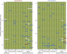
          A total of 149 subjects (mean age 37 years, range 18 to 63 years, 58% women) were recruited. Seropositive infections had been mild with case definition, noncase definition, and asymptomatic disease in 45 (61%), 18 (24%), and 11 (15%), respectively, with 1 person hospitalized (for 2 days). Between seropositive and seronegative groups, there were no differences in cardiac structure (left ventricular volumes, mass, atrial area), function (ejection fraction, global longitudinal shortening, aortic distensibility), tissue characterization (T 1, T 2, extracellular volume fraction mapping, late gadolinium enhancement) or biomarkers (troponin, N-terminal pro–B-type natriuretic peptide). With abnormal defined by the 75 seronegatives (2 SDs from mean, e.g., ejection fraction <54%, septal T 1 >1,072 ms, septal T 2 >52.4 ms), individuals had abnormalities including reduced ejection fraction (n = 2, minimum 50%), T 1 elevation (n = 6), T 2 elevation (n = 9), late gadolinium enhancement (n = 13, median 1%, max 5% of myocardium), biomarker elevation (borderline troponin elevation in 4; all N-terminal pro–B-type natriuretic peptide normal). These were distributed equally between seropositive and seronegative individuals.

          Conclusions

          Cardiovascular abnormalities are no more common in seropositive versus seronegative otherwise healthy, workforce representative individuals 6 months post–mild severe acute respiratory syndrome coronavirus 2 infection.

          Central Illustration

          Related collections

          Most cited references39

          • Record: found
          • Abstract: found
          • Article: not found

          Clinical features of patients infected with 2019 novel coronavirus in Wuhan, China

          Summary Background A recent cluster of pneumonia cases in Wuhan, China, was caused by a novel betacoronavirus, the 2019 novel coronavirus (2019-nCoV). We report the epidemiological, clinical, laboratory, and radiological characteristics and treatment and clinical outcomes of these patients. Methods All patients with suspected 2019-nCoV were admitted to a designated hospital in Wuhan. We prospectively collected and analysed data on patients with laboratory-confirmed 2019-nCoV infection by real-time RT-PCR and next-generation sequencing. Data were obtained with standardised data collection forms shared by WHO and the International Severe Acute Respiratory and Emerging Infection Consortium from electronic medical records. Researchers also directly communicated with patients or their families to ascertain epidemiological and symptom data. Outcomes were also compared between patients who had been admitted to the intensive care unit (ICU) and those who had not. Findings By Jan 2, 2020, 41 admitted hospital patients had been identified as having laboratory-confirmed 2019-nCoV infection. Most of the infected patients were men (30 [73%] of 41); less than half had underlying diseases (13 [32%]), including diabetes (eight [20%]), hypertension (six [15%]), and cardiovascular disease (six [15%]). Median age was 49·0 years (IQR 41·0–58·0). 27 (66%) of 41 patients had been exposed to Huanan seafood market. One family cluster was found. Common symptoms at onset of illness were fever (40 [98%] of 41 patients), cough (31 [76%]), and myalgia or fatigue (18 [44%]); less common symptoms were sputum production (11 [28%] of 39), headache (three [8%] of 38), haemoptysis (two [5%] of 39), and diarrhoea (one [3%] of 38). Dyspnoea developed in 22 (55%) of 40 patients (median time from illness onset to dyspnoea 8·0 days [IQR 5·0–13·0]). 26 (63%) of 41 patients had lymphopenia. All 41 patients had pneumonia with abnormal findings on chest CT. Complications included acute respiratory distress syndrome (12 [29%]), RNAaemia (six [15%]), acute cardiac injury (five [12%]) and secondary infection (four [10%]). 13 (32%) patients were admitted to an ICU and six (15%) died. Compared with non-ICU patients, ICU patients had higher plasma levels of IL2, IL7, IL10, GSCF, IP10, MCP1, MIP1A, and TNFα. Interpretation The 2019-nCoV infection caused clusters of severe respiratory illness similar to severe acute respiratory syndrome coronavirus and was associated with ICU admission and high mortality. Major gaps in our knowledge of the origin, epidemiology, duration of human transmission, and clinical spectrum of disease need fulfilment by future studies. Funding Ministry of Science and Technology, Chinese Academy of Medical Sciences, National Natural Science Foundation of China, and Beijing Municipal Science and Technology Commission.
            Bookmark
            • Record: found
            • Abstract: found
            • Article: not found

            An interactive web-based dashboard to track COVID-19 in real time

            In December, 2019, a local outbreak of pneumonia of initially unknown cause was detected in Wuhan (Hubei, China), and was quickly determined to be caused by a novel coronavirus, 1 namely severe acute respiratory syndrome coronavirus 2 (SARS-CoV-2). The outbreak has since spread to every province of mainland China as well as 27 other countries and regions, with more than 70 000 confirmed cases as of Feb 17, 2020. 2 In response to this ongoing public health emergency, we developed an online interactive dashboard, hosted by the Center for Systems Science and Engineering (CSSE) at Johns Hopkins University, Baltimore, MD, USA, to visualise and track reported cases of coronavirus disease 2019 (COVID-19) in real time. The dashboard, first shared publicly on Jan 22, illustrates the location and number of confirmed COVID-19 cases, deaths, and recoveries for all affected countries. It was developed to provide researchers, public health authorities, and the general public with a user-friendly tool to track the outbreak as it unfolds. All data collected and displayed are made freely available, initially through Google Sheets and now through a GitHub repository, along with the feature layers of the dashboard, which are now included in the Esri Living Atlas. The dashboard reports cases at the province level in China; at the city level in the USA, Australia, and Canada; and at the country level otherwise. During Jan 22–31, all data collection and processing were done manually, and updates were typically done twice a day, morning and night (US Eastern Time). As the outbreak evolved, the manual reporting process became unsustainable; therefore, on Feb 1, we adopted a semi-automated living data stream strategy. Our primary data source is DXY, an online platform run by members of the Chinese medical community, which aggregates local media and government reports to provide cumulative totals of COVID-19 cases in near real time at the province level in China and at the country level otherwise. Every 15 min, the cumulative case counts are updated from DXY for all provinces in China and for other affected countries and regions. For countries and regions outside mainland China (including Hong Kong, Macau, and Taiwan), we found DXY cumulative case counts to frequently lag behind other sources; we therefore manually update these case numbers throughout the day when new cases are identified. To identify new cases, we monitor various Twitter feeds, online news services, and direct communication sent through the dashboard. Before manually updating the dashboard, we confirm the case numbers with regional and local health departments, including the respective centres for disease control and prevention (CDC) of China, Taiwan, and Europe, the Hong Kong Department of Health, the Macau Government, and WHO, as well as city-level and state-level health authorities. For city-level case reports in the USA, Australia, and Canada, which we began reporting on Feb 1, we rely on the US CDC, the government of Canada, the Australian Government Department of Health, and various state or territory health authorities. All manual updates (for countries and regions outside mainland China) are coordinated by a team at Johns Hopkins University. The case data reported on the dashboard aligns with the daily Chinese CDC 3 and WHO situation reports 2 for within and outside of mainland China, respectively (figure ). Furthermore, the dashboard is particularly effective at capturing the timing of the first reported case of COVID-19 in new countries or regions (appendix). With the exception of Australia, Hong Kong, and Italy, the CSSE at Johns Hopkins University has reported newly infected countries ahead of WHO, with Hong Kong and Italy reported within hours of the corresponding WHO situation report. Figure Comparison of COVID-19 case reporting from different sources Daily cumulative case numbers (starting Jan 22, 2020) reported by the Johns Hopkins University Center for Systems Science and Engineering (CSSE), WHO situation reports, and the Chinese Center for Disease Control and Prevention (Chinese CDC) for within (A) and outside (B) mainland China. Given the popularity and impact of the dashboard to date, we plan to continue hosting and managing the tool throughout the entirety of the COVID-19 outbreak and to build out its capabilities to establish a standing tool to monitor and report on future outbreaks. We believe our efforts are crucial to help inform modelling efforts and control measures during the earliest stages of the outbreak.
              Bookmark
              • Record: found
              • Abstract: found
              • Article: found
              Is Open Access

              Features of 20 133 UK patients in hospital with covid-19 using the ISARIC WHO Clinical Characterisation Protocol: prospective observational cohort study

              Abstract Objective To characterise the clinical features of patients admitted to hospital with coronavirus disease 2019 (covid-19) in the United Kingdom during the growth phase of the first wave of this outbreak who were enrolled in the International Severe Acute Respiratory and emerging Infections Consortium (ISARIC) World Health Organization (WHO) Clinical Characterisation Protocol UK (CCP-UK) study, and to explore risk factors associated with mortality in hospital. Design Prospective observational cohort study with rapid data gathering and near real time analysis. Setting 208 acute care hospitals in England, Wales, and Scotland between 6 February and 19 April 2020. A case report form developed by ISARIC and WHO was used to collect clinical data. A minimal follow-up time of two weeks (to 3 May 2020) allowed most patients to complete their hospital admission. Participants 20 133 hospital inpatients with covid-19. Main outcome measures Admission to critical care (high dependency unit or intensive care unit) and mortality in hospital. Results The median age of patients admitted to hospital with covid-19, or with a diagnosis of covid-19 made in hospital, was 73 years (interquartile range 58-82, range 0-104). More men were admitted than women (men 60%, n=12 068; women 40%, n=8065). The median duration of symptoms before admission was 4 days (interquartile range 1-8). The commonest comorbidities were chronic cardiac disease (31%, 5469/17 702), uncomplicated diabetes (21%, 3650/17 599), non-asthmatic chronic pulmonary disease (18%, 3128/17 634), and chronic kidney disease (16%, 2830/17 506); 23% (4161/18 525) had no reported major comorbidity. Overall, 41% (8199/20 133) of patients were discharged alive, 26% (5165/20 133) died, and 34% (6769/20 133) continued to receive care at the reporting date. 17% (3001/18 183) required admission to high dependency or intensive care units; of these, 28% (826/3001) were discharged alive, 32% (958/3001) died, and 41% (1217/3001) continued to receive care at the reporting date. Of those receiving mechanical ventilation, 17% (276/1658) were discharged alive, 37% (618/1658) died, and 46% (764/1658) remained in hospital. Increasing age, male sex, and comorbidities including chronic cardiac disease, non-asthmatic chronic pulmonary disease, chronic kidney disease, liver disease and obesity were associated with higher mortality in hospital. Conclusions ISARIC WHO CCP-UK is a large prospective cohort study of patients in hospital with covid-19. The study continues to enrol at the time of this report. In study participants, mortality was high, independent risk factors were increasing age, male sex, and chronic comorbidity, including obesity. This study has shown the importance of pandemic preparedness and the need to maintain readiness to launch research studies in response to outbreaks. Study registration ISRCTN66726260.
                Bookmark

                Author and article information

                Journal
                JACC Cardiovasc Imaging
                JACC Cardiovasc Imaging
                Jacc. Cardiovascular Imaging
                The Authors. Published by Elsevier on behalf of the American College of Cardiology Foundation.
                1936-878X
                1876-7591
                8 May 2021
                8 May 2021
                Affiliations
                [a ]Barts Heart Centre, Barts Health NHS Trust, West Smithfield, London, United Kingdom
                [b ]Institute of Cardiovascular Science, University College London, London, United Kingdom
                [c ]William Harvey Research Institute, Queen Mary University of London, London, United Kingdom
                [d ]Cardiology Department, University Hospital Centre of Porto, Porto, Portugal
                [e ]Carol Davila University of Medicine and Pharmacy, Bucharest, Romania
                [f ]National Amyloidosis Centre, Division of Medicine, University College London, London, United Kingdom
                [g ]Royal Free London NHS Foundation Trust, Pond Street, London, United Kingdom
                [h ]Cardiology Department, Hospital Prof Doutor Fernando Fonseca Amadora, Portugal
                [i ]Blizard Institute, Queen Mary University of London, London, United Kingdom
                [j ]Division of Infection and Immunity, University College London, London, United Kingdom
                [k ]Imageens, Paris, France
                [l ]Leeds Institute of Cardiovascular and Metabolic Medicine, University of Leeds, and Leeds Teaching Hospitals NHS Trust, Leeds, United Kingdom
                [m ]National Heart, Lung, and Blood Institute, National Institute of Health, Bethesda, Maryland, USA
                [n ]UPMC CMR Center, UPMC, Pittsburgh, Pennsylvania, USA
                Author notes
                [] Address for correspondence: Prof. James Moon, Advanced Cardiac Imaging, St. Bartholomew’s Hospital, West Smithfield, London EC1A 7BE, United Kingdom.
                [∗]

                Drs. Joy and Artico contributed equally to this work and are joint first authors.

                [†]

                Drs. Treibel and Manisty and Prof. Moon contributed equally to this work and are joint last authors.

                Article
                S1936-878X(21)00356-9
                10.1016/j.jcmg.2021.04.011
                8105493
                33975819
                d6328249-6860-4d68-b8b0-0421f01ecf58
                © 2021 The Authors

                Since January 2020 Elsevier has created a COVID-19 resource centre with free information in English and Mandarin on the novel coronavirus COVID-19. The COVID-19 resource centre is hosted on Elsevier Connect, the company's public news and information website. Elsevier hereby grants permission to make all its COVID-19-related research that is available on the COVID-19 resource centre - including this research content - immediately available in PubMed Central and other publicly funded repositories, such as the WHO COVID database with rights for unrestricted research re-use and analyses in any form or by any means with acknowledgement of the original source. These permissions are granted for free by Elsevier for as long as the COVID-19 resource centre remains active.

                History
                : 8 February 2021
                : 8 March 2021
                : 8 April 2021
                Categories
                Original Research

                cardiovascular magnetic resonance,covid-19,late gadolinium enhancement,myocardial edema,myocarditis,sars-cov-2,troponin,cmr, cardiovascular magnetic resonance,covid-19, coronavirus disease-2019,hstnt, high-sensitivity troponin t,lge, late gadolinium enhancement,lv, left ventricular,nt-probnp, n-terminal pro–b-type natriuretic peptide,sars-cov-2, severe acute respiratory syndrome-coronavirus-2

                Comments

                Comment on this article