28
views
0
recommends
+1 Recommend
0 collections
    0
    shares
      • Record: found
      • Abstract: found
      • Article: found
      Is Open Access

      In vitro adhesion properties of Shiga toxin-producing Escherichia coli isolated from cattle, food, and humans

      research-article

      Read this article at

      Bookmark
          There is no author summary for this article yet. Authors can add summaries to their articles on ScienceOpen to make them more accessible to a non-specialist audience.

          Abstract

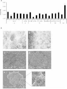
          Shiga toxin-producing Escherichia coli (STEC) are able to cause serious illnesses ranging from diarrhea to hemorrhagic colitis and hemolytic-uremic syndrome (HUS). These bacteria colonize the digestive tract of humans and produce Shiga-toxins, which are considered to be essential for virulence and are crucial in lethal infection. Colon colonization is supposed to be a determinant step in the development of the infection, but the virulence traits that mediate this step are unclear. We analyzed the ability of 256 STEC strains belonging to seropathotype A (the most virulent O157:H7 serotype) to seropathotype E (not involved in human disease) to adhere to HEp-2, HCT-8, and T84 cell lines. Of the 256 STEC tested most (82%) were non-adherent in our assays. The adhesion levels were globally low and were not related to pathogenicity, although the highest levels were associated to O26:H11 and O103:H2 strains of seropathotype B (associated with HUS but less commonly than serotype O157:H7), possessing both the eae and toxB genes.

          Related collections

          Most cited references38

          • Record: found
          • Abstract: found
          • Article: not found

          Pathogenesis and diagnosis of Shiga toxin-producing Escherichia coli infections.

          Since their initial recognition 20 years ago, Shiga toxin-producing Escherichia coli (STEC) strains have emerged as an important cause of serious human gastrointestinal disease, which may result in life-threatening complications such as hemolytic-uremic syndrome. Food-borne outbreaks of STEC disease appear to be increasing and, when mass-produced and mass-distributed foods are concerned, can involve large numbers of people. Development of therapeutic and preventative strategies to combat STEC disease requires a thorough understanding of the mechanisms by which STEC organisms colonize the human intestinal tract and cause local and systemic pathological changes. While our knowledge remains incomplete, recent studies have improved our understanding of these processes, particularly the complex interaction between Shiga toxins and host cells, which is central to the pathogenesis of STEC disease. In addition, several putative accessory virulence factors have been identified and partly characterized. The capacity to limit the scale and severity of STEC disease is also dependent upon rapid and sensitive diagnostic procedures for analysis of human samples and suspect vehicles. The increased application of advanced molecular technologies in clinical laboratories has significantly improved our capacity to diagnose STEC infection early in the course of disease and to detect low levels of environmental contamination. This, in turn, has created a potential window of opportunity for future therapeutic intervention.
            Bookmark
            • Record: found
            • Abstract: found
            • Article: not found

            Association of genomic O island 122 of Escherichia coli EDL 933 with verocytotoxin-producing Escherichia coli seropathotypes that are linked to epidemic and/or serious disease.

            The distribution of EDL 933 O island 122 (OI-122) was investigated in 70 strains of Verocytotoxin-producing Escherichia coli (VTEC) of multiple serotypes that were classified into five "seropathotypes" (A through E) based on the reported occurrence of serotypes in human disease, in outbreaks, and/or in the hemolytic-uremic syndrome (HUS). Seropathotype A comprised 10 serotype O157:H7 and 3 serotype O157:NM strains. Seropathotype B (associated with outbreaks and HUS but less commonly than serotype O157:H7) comprised three strains each of serotypes O26:H11, O103:H2, O111:NM, O121:H19, and O145:NM. Seropathotype C comprised four strains each of serotypes O91:H21 and O113:H21 and eight strains of other serotypes that have been associated with sporadic HUS but not typically with outbreaks. Seropathotype D comprised 14 strains of serotypes that have been associated with diarrhea but not with outbreaks or HUS, and seropathotype E comprised animal VTEC strains of serotypes not implicated in human disease. All strains were tested for four EDL 933 OI-122 virulence genes (Z4321, Z4326, Z4332, and Z4333) by PCR. Negative PCRs were confirmed by Southern hybridization. Overall, 28 (40%) strains contained OI-122 (positive for all four virulence genes), 27 (38.6%) contained an "incomplete" OI-122 (positive for one to three genes), and 15 (21.4%) strains did not contain OI-122. The seropathotype distribution of complete OI-122 was as follows: 100% for seropathotype A, 60% for B, 36% for C, 15% for D, and 0% for E. The differences in the frequency of OI-122 between seropathotypes A, B, and C (associated with HUS) and seropathotypes D and E (not associated with HUS) and between seropathotypes A and B (associated with epidemic disease) and seropathotypes C, D, and E (not associated with epidemic disease) were highly significant (P < 0.0001).
              Bookmark
              • Record: found
              • Abstract: found
              • Article: not found

              Actin accumulation at sites of bacterial adhesion to tissue culture cells: basis of a new diagnostic test for enteropathogenic and enterohemorrhagic Escherichia coli.

              Enteropathogenic Escherichia coli (EPEC) and enterohemorrhagic E. coli (EHEC) adhere to the intestinal mucosa and produce an attaching and effacing (AE) lesion in the brush border microvillous membrane; the AE lesion is characterized by localized destruction of microvilli and intimate attachment of bacteria to the apical enterocyte membrane. A similar lesion is seen when bacteria adhere in vitro to a variety of human tissue culture cell lines. In both cases, dense concentrations of microfilaments are present in the apical cytoplasm beneath attached bacteria. Using a fluorescein-labeled phallotoxin, we have shown that these microfilaments are composed of actin. Cells infected with EPEC and EHEC strains known from electron microscopic studies to produce the AE lesion all exhibited intense spots of fluorescence which corresponded in size and position with each adherent bacterium; cells infected with adherent E. coli strains known not to produce the AE lesion did not produce this striking pattern of fluorescence and were indistinguishable from uninfected control cells. These results indicate that such site-specific concentrations of cytoskeletal actin are characteristic of the AE membrane lesion and can form the basis of a simple, highly sensitive diagnostic test for EPEC and EHEC.
                Bookmark

                Author and article information

                Contributors
                Journal
                Front Microbiol
                Front Microbiol
                Front. Microbiol.
                Frontiers in Microbiology
                Frontiers Media S.A.
                1664-302X
                27 February 2015
                2015
                : 6
                : 156
                Affiliations
                [1] 1Centre de Recherche en Nutrition Humaine Auvergne, M2iSH, ‘Microbes, Intestin, Inflammation et Susceptibilité de l’Hôte’ UMR INSERM/Université d’Auvergne U1071 USC-INRA 2018, Clermont Université – Université d’Auvergne Clermont-Ferrand, France
                [2] 2Centre de Recherche en Nutrition Humaine Auvergne, EA-4678 CIDAM, ‘Conception Ingénierie et Développement de l’Aliment et du Médicament’, Clermont Université – Université d’Auvergne Clermont-Ferrand, France
                [3] 3Unité de Recherche M2iSH, Faculté de Pharmacie, CHU Clermont-Ferrand, Service Bactériologie Mycologie Parasitologie Clermont-Ferrand, France
                Author notes

                Edited by: Christina Maria Joseph Elisabeth Vandenbroucke-Grauls, VU University Medical Center, Netherlands

                Reviewed by: Polly Leung, The Hong Kong Polytechnic University, Hong Kong; Kerry Cooper, United States Department of Agriculture – Agricultural Research Service, USA

                *Correspondence: Valérie Livrelli, Unité de Recherche M2iSH, Faculté de Pharmacie, CHU Clermont-Ferrand, Service Bactériologie Mycologie Parasitologie, 28 Place Henri-Dunant, 63 001 Clermont-Ferrand, France e-mail: valerie.livrelli@ 123456udamail.fr

                Present address: Nathalie Pradel, Aix Marseille Université, IRD, CNRS, Mediterranean Institute of Oceanography, UM 110, 13288 Marseille, France

                This article was submitted to Infectious Diseases, a section of the journal Frontiers in Microbiology.

                Article
                10.3389/fmicb.2015.00156
                4343011
                d5afed7e-b51d-4c85-b004-26d1b7d043c6
                Copyright © 2015 Pradel, Etienne-Mesmin, Thévenot, Cordonnier, Blanquet-Diot and Livrelli.

                This is an open-access article distributed under the terms of the Creative Commons Attribution License (CC BY). The use, distribution or reproduction in other forums is permitted, provided the original author(s) or licensor are credited and that the original publication in this journal is cited, in accordance with accepted academic practice. No use, distribution or reproduction is permitted which does not comply with these terms.

                History
                : 12 December 2014
                : 10 February 2015
                Page count
                Figures: 3, Tables: 2, Equations: 0, References: 44, Pages: 10, Words: 0
                Categories
                Microbiology
                Original Research Article

                Microbiology & Virology
                stec,cell lines adhesion,seropathotype,eae,toxb
                Microbiology & Virology
                stec, cell lines adhesion, seropathotype, eae, toxb

                Comments

                Comment on this article