11
views
0
recommends
+1 Recommend
0 collections
    0
    shares
      • Record: found
      • Abstract: found
      • Article: found
      Is Open Access

      Enterohemorrhagic Escherichia coli pathogenesis: role of Long polar fimbriae in Peyer’s patches interactions

      research-article

      Read this article at

      Bookmark
          There is no author summary for this article yet. Authors can add summaries to their articles on ScienceOpen to make them more accessible to a non-specialist audience.

          Abstract

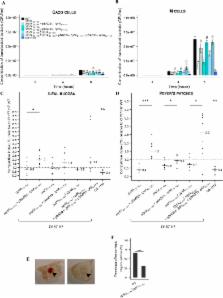
          Enterohemorrhagic Escherichia coli (EHEC) are major food-borne pathogens whose survival and virulence in the human digestive tract remain unclear owing to paucity of relevant models. EHEC interact with the follicle-associated epithelium of Peyer’s patches of the distal ileum and translocate across the intestinal epithelium via M-cells, but the underlying molecular mechanisms are still unknown. Here, we investigated the involvement of Long polar fimbriae (Lpf) in EHEC pathogenesis. Of the 236 strains tested, a significant association was observed between the presence of lpf operons and pathogenicity. In sophisticated in vitro models of the human gastro-intestinal tract, lpf expression was induced during transit through the simulated stomach and small intestine, but not in the colonic compartment. To investigate the involvement of Lpf in EHEC pathogenesis, lpf isogenic mutants and their relative trans-complemented strains were generated. Translocation across M-cells, interactions with murine ileal biopsies containing Peyer’s patches and the number of hemorrhagic lesions were significantly reduced with the lpf mutants compared to the wild-type strain. Complementation of lpf mutants fully restored the wild-type phenotypes. Our results indicate that (i) EHEC might colonize the terminal ileum at the early stages of infection, (ii) Lpf are an important player in the interactions with Peyer’s patches and M-cells, and could contribute to intestinal colonization.

          Related collections

          Most cited references69

          • Record: found
          • Abstract: found
          • Article: not found

          The role of pH in determining the species composition of the human colonic microbiota.

          The pH of the colonic lumen varies with anatomical site and microbial fermentation of dietary residue. We have investigated the impact of mildly acidic pH, which occurs in the proximal colon, on the growth of different species of human colonic bacteria in pure culture and in the complete microbial community. Growth was determined for 33 representative human colonic bacteria at three initial pH values (approximately 5.5, 6.2 and 6.7) in anaerobic YCFA medium, which includes a mixture of short-chain fatty acids (SCFA) with 0.2% glucose as energy source. Representatives of all eight Bacteroides species tested grew poorly at pH 5.5, as did Escherichia coli, whereas 19 of the 23 gram-positive anaerobes tested gave growth rates at pH 5.5 that were at least 50% of those at pH 6.7. Growth inhibition of B. thetaiotaomicron at pH 5.5 was increased by the presence of the SCFA mix (33 mM acetate, 9 mM propionate and 1 mM each of iso-valerate, valerate and iso-butyrate). Analysis of amplified 16S rRNA sequences demonstrated a major pH-driven shift within a human faecal bacterial community in a continuous flow fermentor. Bacteroides spp. accounted for 27% of 16S rRNA sequences detected at pH 5.5, but 86% of sequences at pH 6.7. Conversely, butyrate-producing gram-positive bacteria related to Eubacterium rectale represented 50% of all 16S rRNA sequences at pH 5.5, but were not detected at pH 6.7. Inhibition of the growth of a major group of gram-negative bacteria at mildly acidic pH apparently creates niches that can be exploited by more low pH-tolerant microorganisms.
            Bookmark
            • Record: found
            • Abstract: found
            • Article: not found

            Uptake through glycoprotein 2 of FimH(+) bacteria by M cells initiates mucosal immune response.

            The mucosal immune system forms the largest part of the entire immune system, containing about three-quarters of all lymphocytes and producing grams of secretory IgA daily to protect the mucosal surface from pathogens. To evoke the mucosal immune response, antigens on the mucosal surface must be transported across the epithelial barrier into organized lymphoid structures such as Peyer's patches. This function, called antigen transcytosis, is mediated by specialized epithelial M cells. The molecular mechanisms promoting this antigen uptake, however, are largely unknown. Here we report that glycoprotein 2 (GP2), specifically expressed on the apical plasma membrane of M cells among enterocytes, serves as a transcytotic receptor for mucosal antigens. Recombinant GP2 protein selectively bound a subset of commensal and pathogenic enterobacteria, including Escherichia coli and Salmonella enterica serovar Typhimurium (S. Typhimurium), by recognizing FimH, a component of type I pili on the bacterial outer membrane. Consistently, these bacteria were colocalized with endogenous GP2 on the apical plasma membrane as well as in cytoplasmic vesicles in M cells. Moreover, deficiency of bacterial FimH or host GP2 led to defects in transcytosis of type-I-piliated bacteria through M cells, resulting in an attenuation of antigen-specific immune responses in Peyer's patches. GP2 is therefore a previously unrecognized transcytotic receptor on M cells for type-I-piliated bacteria and is a prerequisite for the mucosal immune response to these bacteria. Given that M cells are considered a promising target for oral vaccination against various infectious diseases, the GP2-dependent transcytotic pathway could provide a new target for the development of M-cell-targeted mucosal vaccines.
              Bookmark
              • Record: found
              • Abstract: found
              • Article: not found

              The risk of the hemolytic-uremic syndrome after antibiotic treatment of Escherichia coli O157:H7 infections.

              Children with gastrointestinal infections caused by Escherichia coli O157:H7 are at risk for the hemolytic-uremic syndrome. Whether antibiotics alter this risk is unknown. We conducted a prospective cohort study of 71 children younger than 10 years of age who had diarrhea caused by E. coli O157:H7 to assess whether antibiotic treatment in these children affects the risk of the hemolytic-uremic syndrome and to assess the influence of confounding factors on this outcome. Estimates of relative risks were adjusted for possible confounding effects with the use of logistic-regression analysis. Among the 71 children, 9 (13 percent) received antibiotics and the hemolytic-uremic syndrome developed in 10 (14 percent). Five of these 10 children had received antibiotics. Factors significantly associated with the hemolytic-uremic syndrome were a higher initial white-cell count (relative risk, 1.3; 95 percent confidence interval, 1.1 to 1.5), evaluation with stool culture soon after the onset of illness (relative risk, 0.3; 95 percent confidence interval, 0.2 to 0.8), and treatment with antibiotics (relative risk, 14.3; 95 percent confidence interval, 2.9 to 70.7). The clinical and laboratory characteristics of the 9 children who received antibiotics and the 62 who did not receive antibiotics were similar. In a multivariate analysis that was adjusted for the initial white-cell count and the day of illness on which stool was obtained for culture, antibiotic administration remained a risk factor for the development of the hemolytic uremic syndrome (relative risk, 17.3; 95 percent confidence interval, 2.2 to 137). Antibiotic treatment of children with E. coli O157:H7 infection increases the risk of the hemolytic-uremic syndrome.
                Bookmark

                Author and article information

                Journal
                Sci Rep
                Sci Rep
                Scientific Reports
                Nature Publishing Group
                2045-2322
                20 March 2017
                2017
                : 7
                : 44655
                Affiliations
                [1 ]Université Clermont Auvergne, Inserm U1071, M2iSH “Microbes, Intestin, Inflammation et Susceptibilité de l’Hôte”, USC-INRA 2018 , F-63000 Clermont-Ferrand, France
                [2 ]Université Clermont Auvergne, MEDIS “Microbiologie Environnement DIgestif Santé” , F-63000 Clermont-Ferrand, France
                [3 ]CHU Clermont-Ferrand, Service de Bactériologie, Parasitologie Mycologie , Clermont-Ferrand, F-63000, France
                Author notes
                [*]

                Present address: Center for Inflammation, Immunity and Infection, Institute for Biomedical Sciences, Georgia State University, Atlanta, GA 30303, USA.

                [†]

                Present address: STLO, “Science et Technologie du Lait et de l’Oeuf ” Agrocampus Ouest, INRA, Rennes, 35042, France.

                [‡]

                Deceased.

                Article
                srep44655
                10.1038/srep44655
                5357955
                28317910
                2d0a4824-bcaa-42b1-b100-fd7a11b235fa
                Copyright © 2017, The Author(s)

                This work is licensed under a Creative Commons Attribution 4.0 International License. The images or other third party material in this article are included in the article’s Creative Commons license, unless indicated otherwise in the credit line; if the material is not included under the Creative Commons license, users will need to obtain permission from the license holder to reproduce the material. To view a copy of this license, visit http://creativecommons.org/licenses/by/4.0/

                History
                : 26 October 2016
                : 13 February 2017
                Categories
                Article

                Uncategorized
                Uncategorized

                Comments

                Comment on this article