3
views
0
recommends
+1 Recommend
0 collections
    0
    shares
      • Record: found
      • Abstract: found
      • Article: found
      Is Open Access

      Adherens junction proteins on the move—From the membrane to the nucleus in intestinal diseases

      review-article

      Read this article at

      Bookmark
          There is no author summary for this article yet. Authors can add summaries to their articles on ScienceOpen to make them more accessible to a non-specialist audience.

          Abstract

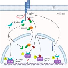
          The function and structure of the mammalian epithelial cell layer is maintained by distinct intercellular adhesion complexes including adherens junctions (AJs), tight junctions, and desmosomes. The AJ is most integral for stabilizing cell-cell adhesion and conserving the structural integrity of epithelial tissues. AJs are comprised of the transmembrane protein E-cadherin and cytoplasmic catenin cofactors (α, β, γ, and p120-catenin). One organ where malfunction of AJ is a major contributor to disease states is the mammalian intestine. In the intestine, cell-cell adhesion complexes work synergistically to maintain structural integrity and homeostasis of the epithelium and prevent its malfunction. Consequently, when AJ integrity is compromised in the intestinal epithelium, the ensuing homeostatic disruption leads to diseases such as inflammatory bowel disease and colorectal carcinoma. In addition to their function at the plasma membrane, protein components of AJs also have nuclear functions and are thus implicated in regulating gene expression and intracellular signaling. Within the nucleus, AJ proteins have been shown to interact with transcription factors such as TCF/LEF and Kaiso ( ZBTB33), which converge on the canonical Wnt signaling pathway. The multifaceted nature of AJ proteins highlights their complexity in modulating homeostasis and emphasizes the importance of their subcellular localization and expression in the mammalian intestine. In this review, we summarize the nuclear roles of AJ proteins in intestinal tissues; their interactions with transcription factors and how this leads to crosstalk with canonical Wnt signaling; and how nuclear AJ proteins are implicated in intestinal homeostasis and disease.

          Related collections

          Most cited references165

          • Record: found
          • Abstract: found
          • Article: not found

          Intestinal epithelial cells: regulators of barrier function and immune homeostasis.

          The abundance of innate and adaptive immune cells that reside together with trillions of beneficial commensal microorganisms in the mammalian gastrointestinal tract requires barrier and regulatory mechanisms that conserve host-microbial interactions and tissue homeostasis. This homeostasis depends on the diverse functions of intestinal epithelial cells (IECs), which include the physical segregation of commensal bacteria and the integration of microbial signals. Hence, IECs are crucial mediators of intestinal homeostasis that enable the establishment of an immunological environment permissive to colonization by commensal bacteria. In this Review, we provide a comprehensive overview of how IECs maintain host-commensal microbial relationships and immune cell homeostasis in the intestine.
            Bookmark
            • Record: found
            • Abstract: found
            • Article: not found

            The many faces and functions of β-catenin.

            β-Catenin (Armadillo in Drosophila) is a multitasking and evolutionary conserved molecule that in metazoans exerts a crucial role in a multitude of developmental and homeostatic processes. More specifically, β-catenin is an integral structural component of cadherin-based adherens junctions, and the key nuclear effector of canonical Wnt signalling in the nucleus. Imbalance in the structural and signalling properties of β-catenin often results in disease and deregulated growth connected to cancer and metastasis. Intense research into the life of β-catenin has revealed a complex picture. Here, we try to capture the state of the art: we try to summarize and make some sense of the processes that regulate β-catenin, as well as the plethora of β-catenin binding partners. One focus will be the interaction of β-catenin with different transcription factors and the potential implications of these interactions for direct cross-talk between β-catenin and non-Wnt signalling pathways.
              Bookmark
              • Record: found
              • Abstract: found
              • Article: not found

              Axin, a negative regulator of the Wnt signaling pathway, forms a complex with GSK-3beta and beta-catenin and promotes GSK-3beta-dependent phosphorylation of beta-catenin.

              Glycogen synthase kinase-3 (GSK-3) mediates epidermal growth factor, insulin and Wnt signals to various downstream events such as glycogen metabolism, gene expression, proliferation and differentiation. We have isolated here a GSK-3beta-interacting protein from a rat brain cDNA library using a yeast two-hybrid method. This protein consists of 832 amino acids and possesses Regulators of G protein Signaling (RGS) and dishevelled (Dsh) homologous domains in its N- and C-terminal regions, respectively. The predicted amino acid sequence of this GSK-3beta-interacting protein shows 94% identity with mouse Axin, which recently has been identified as a negative regulator of the Wnt signaling pathway; therefore, we termed this protein rAxin (rat Axin). rAxin interacted directly with, and was phosphorylated by, GSK-3beta. rAxin also interacted directly with the armadillo repeats of beta-catenin. The binding site of rAxin for GSK-3beta was distinct from the beta-catenin-binding site, and these three proteins formed a ternary complex. Furthermore, rAxin promoted GSK-3beta-dependent phosphorylation of beta-catenin. These results suggest that rAxin negatively regulates the Wnt signaling pathway by interacting with GSK-3beta and beta-catenin and mediating the signal from GSK-3beta to beta-catenin.
                Bookmark

                Author and article information

                Contributors
                Journal
                Front Cell Dev Biol
                Front Cell Dev Biol
                Front. Cell Dev. Biol.
                Frontiers in Cell and Developmental Biology
                Frontiers Media S.A.
                2296-634X
                05 October 2022
                2022
                : 10
                : 998373
                Affiliations
                Department of Biology , McMaster University , Hamilton, ON, Canada
                Author notes

                Edited by: Antonis Kourtidis, Medical University of South Carolina, United States

                Reviewed by: Andrei Ivanov, Cleveland Clinic, United States

                Orest William Blaschuk, Zonula Inc., Canada

                René-Marc Mège, Centre National de la Recherche Scientifique (CNRS), France

                *Correspondence: Juliet M. Daniel, danielj@ 123456mcmaster.ca
                [ † ]

                Present Address: Shaiya C. Robinson, School of Interdisciplinary Science, McMaster University, Hamilton, ON, Canada

                [ ‡ ]

                These authors share first authorship

                This article was submitted to Cell Adhesion and Migration, a section of the journal Frontiers in Cell and Developmental Biology

                Article
                998373
                10.3389/fcell.2022.998373
                9581404
                36274850
                d4d7f9db-2c4d-4882-879c-cac73576bb08
                Copyright © 2022 Lessey, Robinson, Chaudhary and Daniel.

                This is an open-access article distributed under the terms of the Creative Commons Attribution License (CC BY). The use, distribution or reproduction in other forums is permitted, provided the original author(s) and the copyright owner(s) are credited and that the original publication in this journal is cited, in accordance with accepted academic practice. No use, distribution or reproduction is permitted which does not comply with these terms.

                History
                : 19 July 2022
                : 29 August 2022
                Funding
                Funded by: Natural Sciences and Engineering Research Council of Canada , doi 10.13039/501100000038;
                Funded by: Cancer Research Society , doi 10.13039/100009326;
                Categories
                Cell and Developmental Biology
                Review

                adherens junction,e-cadherin,catenins,kaiso,wnt signaling,inflammatory bowel disease,colon cancer

                Comments

                Comment on this article