26
views
0
recommends
+1 Recommend
0 collections
    0
    shares
      • Record: found
      • Abstract: found
      • Article: found
      Is Open Access

      Interferon-Alpha Promotes Th1 Response and Epithelial Apoptosis via Inflammasome Activation in Human Intestinal Mucosa

      research-article

      Read this article at

      Bookmark
          There is no author summary for this article yet. Authors can add summaries to their articles on ScienceOpen to make them more accessible to a non-specialist audience.

          Abstract

          Backgound & Aims

          Several lines of investigation suggest that interferon (IFN) alpha can alter human intestinal mucosa homeostasis. These include the endogenous production of IFN alpha in celiac disease or inflammatory bowel diseases, as well as the occurrence of intestinal side effects of exogenous IFN alpha used as a therapeutic tool. Here, we present an ex vivo translational approach to investigate the effects of IFN alpha on the human normal intestinal mucosa, as well as its underlying mechanisms.

          Methods

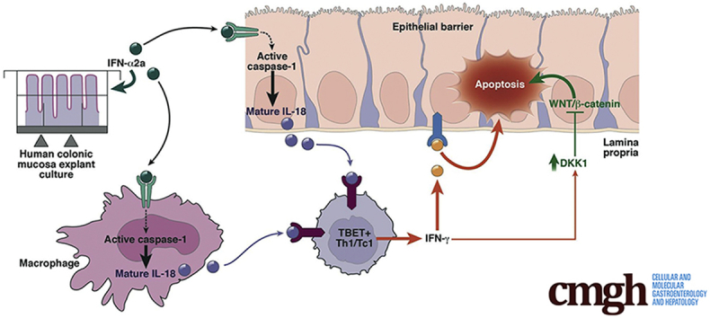
          Human normal colonic mucosa explants were cultured in the presence or absence of IFN alpha 2a. Epithelial homeostasis was assessed using the immunohistochemical marker of apoptosis M30. The Wnt inhibitor Dickkopf-Homolog-1 (DKK1) was assayed in the supernatants by enzyme-linked immunosorbent assay. Activation of the inflammasome (caspase-1/interleukin [IL]18) and of a Th1 response was determined by in situ detection of active caspase-1, as well as by measurement of mature IL18 production and the prototype Th1 cytokine IFN gamma by enzyme-linked immunosorbent assay. In addition, mechanistic studies were performed using the specific caspase-1 inhibitor Tyr-Val-Ala-Asp(OMe)-fluoromethylketone (YVAD-FMK), IL18-binding protein, neutralizing anti–IFN gamma, and anti-DKK1 antibodies.

          Results

          IFN alpha 2a elicited a rapid (24 hours) disruption of surface and crypt colonic epithelial cells via apoptosis that was variable in intensity among the 20 individuals studied. This apoptotic effect was dependent on the initiation of an IFN gamma response elicited by resident T box expressed in T cells–positive lamina propria cells. Both apoptosis and Th1 response were subordinated to active caspase-1 and IL18 production. Finally, neutralization of IFN gamma–induced DKK1 partially protected against IFN alpha–induced epithelial apoptosis.

          Conclusions

          By using an ex vivo model, we show an interindividual heterogeneity of IFN alpha effects. We show that IFN alpha is able to disrupt both epithelial and immune homeostasis in the human intestine, by activation of an innate immunity platform, the inflammasome, which drives a Th1 response and leads to epithelial barrier disruption.

          Graphical abstract

          Related collections

          Most cited references33

          • Record: found
          • Abstract: found
          • Article: not found

          The inflammasomes: guardians of the body.

          The innate immune system relies on its capacity to rapidly detect invading pathogenic microbes as foreign and to eliminate them. The discovery of Toll-like receptors (TLRs) provided a class of membrane receptors that sense extracellular microbes and trigger antipathogen signaling cascades. More recently, intracellular microbial sensors have been identified, including NOD-like receptors (NLRs). Some of the NLRs also sense nonmicrobial danger signals and form large cytoplasmic complexes called inflammasomes that link the sensing of microbial products and metabolic stress to the proteolytic activation of the proinflammatory cytokines IL-1beta and IL-18. The NALP3 inflammasome has been associated with several autoinflammatory conditions including gout. Likewise, the NALP3 inflammasome is a crucial element in the adjuvant effect of aluminum and can direct a humoral adaptive immune response. In this review, we discuss the role of NLRs, and in particular the inflammasomes, in the recognition of microbial and danger components and the role they play in health and disease.
            Bookmark
            • Record: found
            • Abstract: found
            • Article: not found

            Proinflammatory cytokines disrupt epithelial barrier function by apoptosis-independent mechanisms.

            It is well known that inflammatory conditions of the intestinal mucosa result in compromised barrier function. Inflammation is characterized by an influx into the mucosa of immune cells that influence epithelial function by releasing proinflammatory cytokines such as IFN-gamma and TNF-alpha. Mucosal barrier function is regulated by the epithelial apical junctional complex (AJC) consisting of the tight junction and the adherens junction. Since the AJC regulates barrier function, we analyzed the influence of IFN-gamma and TNF-alpha on its structure/function and determined the contribution of apoptosis to this process using a model intestinal epithelial cell line, T84, and IFN-gamma and TNF-alpha. AJC structure/function was analyzed by confocal microscopy, biochemical analysis, and physiologic measurement of epithelial gate/fence function. Apoptosis was monitored by determining cytokeratin 18 cleavage and caspase-3 activation. IFN-gamma induced time-dependent disruptions in epithelial gate function that were potentiated by coincubation with TNF-alpha. Tight junction fence function was somewhat disrupted. Cytokine treatment was associated with internalization of AJC transmembrane proteins, junction adhesion molecule 1, occludin, and claudin-1/4 with minimal effects on the cytoplasmic plaque protein zonula occludens 1. Detergent solubility profiles of junction adhesion molecule 1 and E-cadherin and their affiliation with "raft-like" membrane microdomains were modified by these cytokines. Inhibition of cytokine-induced apoptosis did not block induced permeability defects; further emphasizing their primary influence on the epithelial AJC structure and barrier function. Our findings for the first time clearly separate the proapoptotic effects of IFN-gamma and TNF-alpha from their abilities to disrupt barrier function.
              Bookmark
              • Record: found
              • Abstract: found
              • Article: not found

              Interferon-gamma regulates intestinal epithelial homeostasis through converging beta-catenin signaling pathways.

              Inflammatory cytokines have been proposed to regulate epithelial homeostasis during intestinal inflammation. We report here that interferon-gamma (IFN-gamma) regulates the crucial homeostatic functions of cell proliferation and apoptosis through serine-threonine protein kinase AKT-beta-catenin and Wingless-Int (Wnt)-beta-catenin signaling pathways. Short-term exposure of intestinal epithelial cells to IFN-gamma resulted in activation of beta-catenin through AKT, followed by induction of the secreted Wnt inhibitor Dkk1. Consequently, we observed an increase in Dkk1-mediated apoptosis upon extended IFN-gamma treatment and reduced proliferation through depletion of the Wnt coreceptor LRP6. These effects were enhanced by tumor necrosis factor-alpha (TNF-alpha), suggesting synergism between the two cytokines. Consistent with these results, colitis in vivo was associated with decreased beta-catenin-T cell factor (TCF) signaling, loss of plasma membrane-associated LRP6, and reduced epithelial cell proliferation. Proliferation was partially restored in IFN-gamma-deficient mice. Thus, we propose that IFN-gamma regulates intestinal epithelial homeostasis by sequential regulation of converging beta-catenin signaling pathways.
                Bookmark

                Author and article information

                Contributors
                Journal
                Cell Mol Gastroenterol Hepatol
                Cell Mol Gastroenterol Hepatol
                Cellular and Molecular Gastroenterology and Hepatology
                Elsevier
                2352-345X
                20 September 2016
                January 2017
                20 September 2016
                : 3
                : 1
                : 72-81
                Affiliations
                [1 ]EA4273 Biometadys, Faculté de Médecine, Université de Nantes, Nantes, France
                [2 ]Center for Research in Transplantation and Immunology, INSERM UMR1064, Nantes, France
                [3 ]Service d’Hématologie Clinique, Centre Hospitalo-Universitaire de Nantes, Nantes, France
                [6 ]Clinique de Chirurgie Digestive et Endocrinienne, Centre Hospitalo-Universitaire de Nantes, Nantes, France
                [7 ]Service d’Anatomie et Cytologie Pathologiques, Centre Hospitalo-Universitaire de Nantes, Nantes, France
                [4 ]Centre de Recherche Saint-Antoine, INSERM UMR 938, Paris, France
                [5 ]Service d'Hématologie Clinique et de Thérapie Cellulaire, Hôpital Saint Antoine, Assistance Publique - Hôpitaux de Paris, Paris, France
                Author notes
                [] Correspondence Address correspondence to: Anne Jarry, PhD, or Céline Bossard, MD, PhD, EA4273 Biometadys, Faculté de Médecine, 1 Rue Gaston Veil, 44035 Nantes Cedex 1, France. fax: (33) 2-40-08-47-02.EA4273 Biometadys, Faculté de Médecine1 Rue Gaston Veil, 44035 Nantes Cedex 1France anne.jarry@ 123456univ-nantes.fr celine.bossard@ 123456chu-nantes.fr
                Article
                S2352-345X(16)30108-4
                10.1016/j.jcmgh.2016.09.007
                5247398
                28174758
                d4d167cf-72f5-4baf-b51a-b4898a3440ec
                © 2017 The Authors

                This is an open access article under the CC BY-NC-ND license (http://creativecommons.org/licenses/by-nc-nd/4.0/).

                History
                : 18 March 2016
                : 12 September 2016
                Categories
                Original Research

                caspase-1,mucosal innate and adaptive immunity,roferon,dkk1,dkk1, dickkopf-homolog-1,elisa, enzyme-linked immunosorbent assay,flica, fluorescent-labeled inhibitor of caspases,ifn, interferon,il, interleukin,il18-bp, interleukin 18-binding protein,t-bet, t box expressed in t cells,tc1, cytotoxic t cells type 1,th, t-helper,3d, 3-dimensional,yvad-fmk, tyr-val-ala-asp(ome)-fluoromethylketone

                Comments

                Comment on this article