10
views
0
recommends
+1 Recommend
0 collections
    0
    shares
      • Record: found
      • Abstract: found
      • Article: found
      Is Open Access

      Shaped 3D microcarriers for adherent cell culture and analysis

      research-article

      Read this article at

      Bookmark
          There is no author summary for this article yet. Authors can add summaries to their articles on ScienceOpen to make them more accessible to a non-specialist audience.

          Abstract

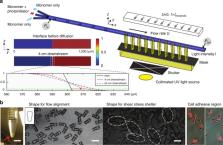
          Standard tissue culture of adherent cells is known to poorly replicate physiology and often entails suspending cells in solution for analysis and sorting, which modulates protein expression and eliminates intercellular connections. To allow adherent culture and processing in flow, we present 3D-shaped hydrogel cell microcarriers, which are designed with a recessed nook in a first dimension to provide a tunable shear-stress shelter for cell growth, and a dumbbell shape in an orthogonal direction to allow for self-alignment in a confined flow, important for processing in flow and imaging flow cytometry. We designed a method to rapidly design, using the genetic algorithm, and manufacture the microcarriers at scale using a transient liquid molding optofluidic approach. The ability to precisely engineer the microcarriers solves fundamental challenges with shear-stress-induced cell damage during liquid-handling, and is poised to enable adherent cell culture, in-flow analysis, and sorting in a single format.

          Adherent cells: microcarriers for flow cytometry

          A new microcarrier for adherent cells is demonstrated which allows for accurate flow cytometry and high-speed imaging without risk of flow-induced damage. Microcarriers are attractive for accelerated cell culture, passaging and analysis, but they must be designed to promote cell growth and analysis without flow-induced cell damage. A team led by Dino Di Carlo at University of California, Los Angeles now report a 3D-shaped microparticle that features a region of extracellular matrix for cell adhesion and culture physically protected from shear flow. Key to the design is the intersection of two 2D patterns, leading to a shape which can align with flow inside the channel during cytometry, and also provides a cut-away region to protect cells during culture. These microcarriers may facilitate high-speed adherent cell screening for applications such as drug discovery.

          Related collections

          Most cited references21

          • Record: found
          • Abstract: found
          • Article: not found

          Generation of human induced pluripotent stem cells from dermal fibroblasts.

          The generation of patient-specific pluripotent stem cells has the potential to accelerate the implementation of stem cells for clinical treatment of degenerative diseases. Technologies including somatic cell nuclear transfer and cell fusion might generate such cells but are hindered by issues that might prevent them from being used clinically. Here, we describe methods to use dermal fibroblasts easily obtained from an individual human to generate human induced pluripotent stem (iPS) cells by ectopic expression of the defined transcription factors KLF4, OCT4, SOX2, and C-MYC. The resultant cell lines are morphologically indistinguishable from human embryonic stem cells (HESC) generated from the inner cell mass of a human preimplantation embryo. Consistent with these observations, human iPS cells share a nearly identical gene-expression profile with two established HESC lines. Importantly, DNA fingerprinting indicates that the human iPS cells were derived from the donor material and are not a result of contamination. Karyotypic analyses demonstrate that reprogramming of human cells by defined factors does not induce, or require, chromosomal abnormalities. Finally, we provide evidence that human iPS cells can be induced to differentiate along lineages representative of the three embryonic germ layers indicating the pluripotency of these cells. Our findings are an important step toward manipulating somatic human cells to generate an unlimited supply of patient-specific pluripotent stem cells. In the future, the use of defined factors to change cell fate may be the key to routine nuclear reprogramming of human somatic cells.
            Bookmark
            • Record: found
            • Abstract: found
            • Article: not found

            Directed assembly of cell-laden microgels for fabrication of 3D tissue constructs.

            We present a bottom-up approach to direct the assembly of cell-laden microgels to generate tissue constructs with tunable microarchitecture and complexity. This assembly process is driven by the tendency of multiphase liquid-liquid systems to minimize the surface area and the resulting surface free energy between the phases. We demonstrate that shape-controlled microgels spontaneously assemble within multiphase reactor systems into predetermined geometric configurations. Furthermore, we characterize the parameters that influence the assembly process, such as external energy input, surface tension, and microgel dimensions. Finally, we show that multicomponent cell-laden constructs could be generated by assembling microgel building blocks and performing a secondary cross-linking reaction. This bottom-up approach for the directed assembly of cell-laden microgels provides a powerful and highly scalable approach to form biomimetic 3D tissue constructs and opens a paradigm for directing the assembly of mesoscale materials.
              Bookmark
              • Record: found
              • Abstract: found
              • Article: not found

              A high throughput platform for stem cell-niche co-cultures and downstream gene expression analysis

              Stem cells reside in “niches”, where support cells provide signaling critical for tissue renewal. Culture methods mimic niche conditions and support the growth of stem cells in vitro. However, current functional assays preclude statistically meaningful studies of clonal stem cells, stem cell-niche interactions, and genetic analysis of single cells and their organoid progeny. Here, we describe a “microraft array” (MRA) that facilitates high-throughput clonogenic culture and computational identification of single intestinal stem cells (ISCs) and niche cells co-cultures. We use MRAs to demonstrate that Paneth cells, a known ISC niche component, enhance organoid formation in a contact-dependent manner. MRAs facilitate retrieval of early enteroids for qPCR to correlate functional properties, such as enteroid morphology, with differences in gene expression. MRAs have broad applicability to assaying stem cell-niche interactions and organoid development, and serve as a high-throughput culture platform to interrogate gene expression at early stages of stem cell fate choices.
                Bookmark

                Author and article information

                Contributors
                dicarlo@ucla.edu
                Journal
                Microsyst Nanoeng
                Microsyst Nanoeng
                Microsystems & Nanoengineering
                Nature Publishing Group UK (London )
                2096-1030
                2055-7434
                13 August 2018
                13 August 2018
                2018
                : 4
                : 21
                Affiliations
                [1 ]ISNI 0000 0000 9632 6718, GRID grid.19006.3e, Department of Bioengineering, , University of California, ; Los Angeles, CA USA
                [2 ]ISNI 0000 0000 9632 6718, GRID grid.19006.3e, Department of Mechanical and Aerospace Engineering, , University of California, ; Los Angeles, CA USA
                [3 ]ISNI 0000 0000 9632 6718, GRID grid.19006.3e, California NanoSystems Institute, , University of California, ; Los Angeles, CA USA
                [4 ]ISNI 0000 0000 9632 6718, GRID grid.19006.3e, Jonsson Comprehensive Cancer Center, , University of California, ; Los Angeles, CA USA
                [5 ]ISNI 0000 0004 1936 7312, GRID grid.34421.30, Department of Mechanical Engineering, , Iowa State University, ; Ames, IA USA
                Article
                20
                10.1038/s41378-018-0020-7
                6220171
                31057909
                d4582964-b53a-4f57-82c5-c2b35fe9c84c
                © The Author(s) 2018

                Open Access This article is licensed under a Creative Commons Attribution 4.0 International License, which permits use, sharing, adaptation, distribution and reproduction in any medium or format, as long as you give appropriate credit to the original author(s) and the source, provide a link to the Creative Commons license, and indicate if changes were made. The images or other third party material in this article are included in the article’s Creative Commons license, unless indicated otherwise in a credit line to the material. If material is not included in the article’s Creative Commons license and your intended use is not permitted by statutory regulation or exceeds the permitted use, you will need to obtain permission directly from the copyright holder. To view a copy of this license, visit http://creativecommons.org/licenses/by/4.0/.

                History
                : 9 January 2018
                : 30 April 2018
                Funding
                Funded by: FundRef https://doi.org/10.13039/100000001, National Science Foundation (NSF);
                Award ID: NSF 1306866, NSF 1306866
                Award Recipient :
                Funded by: Presidential Early Career Award for Scientists and Engineers (N00014-16-1-2997)
                Categories
                Article
                Custom metadata
                © The Author(s) 2018

                Comments

                Comment on this article