27
views
0
recommends
+1 Recommend
0 collections
    0
    shares
      • Record: found
      • Abstract: found
      • Article: found
      Is Open Access

      Offline impact of transcranial focused ultrasound on cortical activation in primates

      research-article

      Read this article at

      Bookmark
          There is no author summary for this article yet. Authors can add summaries to their articles on ScienceOpen to make them more accessible to a non-specialist audience.

          Abstract

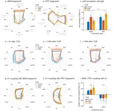
          To understand brain circuits it is necessary both to record and manipulate their activity. Transcranial ultrasound stimulation (TUS) is a promising non-invasive brain stimulation technique. To date, investigations report short-lived neuromodulatory effects, but to deliver on its full potential for research and therapy, ultrasound protocols are required that induce longer-lasting ‘offline’ changes. Here, we present a TUS protocol that modulates brain activation in macaques for more than one hour after 40 s of stimulation, while circumventing auditory confounds. Normally activity in brain areas reflects activity in interconnected regions but TUS caused stimulated areas to interact more selectively with the rest of the brain. In a within-subject design, we observe regionally specific TUS effects for two medial frontal brain regions – supplementary motor area and frontal polar cortex. Independently of these site-specific effects, TUS also induced signal changes in the meningeal compartment. TUS effects were temporary and not associated with microstructural changes.

          eLife digest

          Ultrasound is well known for making visible what is hidden, for example, when giving parents a glimpse of their child before birth. But researchers are now using these high-frequency sound waves – beyond the range of human hearing – for a wholly different purpose: to manipulate the activity of the brain. Conventional brain stimulation techniques use electric currents or magnetic fields to alter brain activity. These techniques, however, have limitations. They can only reach the surface of the brain and are not particularly precise. By contrast, beams of ultrasound can be focused at a millimetre scale, even deep within the brain. Ultrasound thus has the potential to provide new insights into how the brain works.

          Most studies of ultrasound stimulation have looked at what happens to the brain during the stimulation itself. But could ultrasound also induce longer-lasting changes in brain activity? Changes that persist after the stimulation has ended would be valuable for research. They would also make it more likely that we could use ultrasound to treat brain disorders by changing brain activity.

          Verhagen, Gallea et al. used a brain scanner to measure brain activity in macaque monkeys after ultrasound stimulation. The results showed that 40 seconds of repetitive ultrasound changed brain activity for up to two hours. Ultrasound caused the stimulated brain area to interact more selectively with the rest of the brain. Notably, only the stimulated area changed its activity in this way. This helps rule out the possibility that the changes reflect non-specific effects. If the monkeys had been able to hear the ultrasound, for example, it would have changed the activity of the parts of the brain related to hearing. Most important of all, the changes were reversible and did not harm the brain.

          The results of Verhagen, Gallea et al. show that repetitive ultrasound can induce long-lasting alterations in brain activity. It can target areas deep within the brain, including those that are out of reach with other techniques. If this procedure also shows longer-lasting effects in people, it could yield valuable insights into the links between brain and behaviour. It could also help us develop new treatments for neurological and psychiatric disorders.

          Related collections

          Most cited references57

          • Record: found
          • Abstract: found
          • Article: not found

          Situating the default-mode network along a principal gradient of macroscale cortical organization.

          Understanding how the structure of cognition arises from the topographical organization of the cortex is a primary goal in neuroscience. Previous work has described local functional gradients extending from perceptual and motor regions to cortical areas representing more abstract functions, but an overarching framework for the association between structure and function is still lacking. Here, we show that the principal gradient revealed by the decomposition of connectivity data in humans and the macaque monkey is anchored by, at one end, regions serving primary sensory/motor functions and at the other end, transmodal regions that, in humans, are known as the default-mode network (DMN). These DMN regions exhibit the greatest geodesic distance along the cortical surface-and are precisely equidistant-from primary sensory/motor morphological landmarks. The principal gradient also provides an organizing spatial framework for multiple large-scale networks and characterizes a spectrum from unimodal to heteromodal activity in a functional metaanalysis. Together, these observations provide a characterization of the topographical organization of cortex and indicate that the role of the DMN in cognition might arise from its position at one extreme of a hierarchy, allowing it to process transmodal information that is unrelated to immediate sensory input.
            Bookmark
            • Record: found
            • Abstract: not found
            • Article: not found

            Analysis of tissue and arterial blood temperatures in the resting human forearm.

            H H PENNES (1948)
              Bookmark
              • Record: found
              • Abstract: found
              • Article: not found

              Transcranial pulsed ultrasound stimulates intact brain circuits.

              Electromagnetic-based methods of stimulating brain activity require invasive procedures or have other limitations. Deep-brain stimulation requires surgically implanted electrodes. Transcranial magnetic stimulation does not require surgery, but suffers from low spatial resolution. Optogenetic-based approaches have unrivaled spatial precision, but require genetic manipulation. In search of a potential solution to these limitations, we began investigating the influence of transcranial pulsed ultrasound on neuronal activity in the intact mouse brain. In motor cortex, ultrasound-stimulated neuronal activity was sufficient to evoke motor behaviors. Deeper in subcortical circuits, we used targeted transcranial ultrasound to stimulate neuronal activity and synchronous oscillations in the intact hippocampus. We found that ultrasound triggers TTX-sensitive neuronal activity in the absence of a rise in brain temperature (<0.01 degrees C). Here, we also report that transcranial pulsed ultrasound for intact brain circuit stimulation has a lateral spatial resolution of approximately 2 mm and does not require exogenous factors or surgical invasion. Copyright 2010 Elsevier Inc. All rights reserved.
                Bookmark

                Author and article information

                Contributors
                Role: Senior Editor
                Role: Reviewing Editor
                Journal
                eLife
                Elife
                eLife
                eLife
                eLife Sciences Publications, Ltd
                2050-084X
                12 February 2019
                2019
                : 8
                : e40541
                Affiliations
                [1 ]deptWellcome Centre for Integrative Neuroimaging (WIN), Department of Experimental Psychology University of Oxford OxfordUnited Kingdom
                [2 ]deptWellcome Centre for Integrative Neuroimaging (WIN), Centre for Functional MRI of the Brain (FMRIB), Nuffield Department of Clinical Neurosciences John Radcliffe Hospital, University of Oxford OxfordUnited Kingdom
                [3 ]deptInstitute du Cerveau et de la Moelle épinière (ICM), Centre for NeuroImaging Research (CENIR) Inserm U 1127, CNRS UMR 7225, Sorbonne Université ParisFrance
                [4 ]deptPhysics for Medicine Paris Inserm, ESPCI Paris, CNRS, PSL Research University, Université Paris Diderot, Sorbonne Paris Cité ParisFrance
                [5 ]deptDepartment of Physiology, Anatomy and Genetics University of Oxford OxfordUnited Kingdom
                [6 ]deptDonders Institute for Brain, Cognition and Behaviour Radboud University Nijmegen NijmegenThe Netherlands
                [7 ]deptInstitute du Cerveau et de la Moelle épinière (ICM) UMRS 975 INSERM, CNRS 7225, UMPC ParisFrance
                [8 ]deptPhysics for Medicine Paris Inserm, ESPCI Paris, CNRS, PSL Research University ParisFrance
                University of Pennsylvania United States
                University College London United Kingdom
                University College London United Kingdom
                University of Western Ontario
                City College of New York United States
                Author notes
                [†]

                These authors contributed equally to this work.

                [‡]

                These authors also contributed equally to this work.

                Author information
                http://orcid.org/0000-0003-3207-7929
                http://orcid.org/0000-0003-1653-5969
                http://orcid.org/0000-0001-6378-9158
                http://orcid.org/0000-0002-5156-9833
                http://orcid.org/0000-0001-7119-9350
                http://orcid.org/0000-0002-4721-7376
                http://orcid.org/0000-0003-2644-3945
                http://orcid.org/0000-0002-7878-0209
                Article
                40541
                10.7554/eLife.40541
                6372282
                30747105
                d1b03f6e-1d86-449c-bf1c-f23eb7d566b9
                © 2019, Verhagen et al

                This article is distributed under the terms of the Creative Commons Attribution License, which permits unrestricted use and redistribution provided that the original author and source are credited.

                History
                : 28 July 2018
                : 26 January 2019
                Funding
                Funded by: FundRef http://dx.doi.org/10.13039/100004440, Wellcome;
                Award ID: 203139/Z/16/Z
                Award Recipient :
                Funded by: FundRef http://dx.doi.org/10.13039/100004440, Wellcome;
                Award ID: WT100973AIA
                Award Recipient :
                Funded by: Bettencourt Schueller Foundation;
                Award Recipient :
                Funded by: Agence Nationale de la Recherche;
                Award ID: ANR-10-EQPX-15
                Award Recipient :
                Funded by: FundRef http://dx.doi.org/10.13039/100004440, Wellcome;
                Award ID: 105238/Z/14/Z
                Award Recipient :
                Funded by: Medical Research Council;
                Award ID: MR/P024955/1
                Award Recipient :
                Funded by: FundRef http://dx.doi.org/10.13039/100004440, Wellcome;
                Award ID: 103184/Z/13/Z
                Award Recipient :
                Funded by: Biotechnology and Biological Sciences Research Council;
                Award ID: BB/N019814/1
                Award Recipient :
                Funded by: Nederlandse Organisatie voor Wetenschappelijk Onderzoek;
                Award ID: 452-13-015
                Award Recipient :
                Funded by: FundRef http://dx.doi.org/10.13039/501100000265, Medical Research Council;
                Award ID: G0902373
                Award Recipient :
                Funded by: FundRef http://dx.doi.org/10.13039/100004440, Wellcome;
                Award ID: 105651/Z/14/Z
                Award Recipient :
                The funders had no role in study design, data collection and interpretation, or the decision to submit the work for publication.
                Categories
                Research Article
                Neuroscience
                Custom metadata
                A new application of transcranial ultrasound safely modulates brain activation in primates for up to 2 hours after 40 seconds of stimulation.

                Life sciences
                brain stimulation,transcranial ultrasound stimulation,neuroimaging,resting-state fmri,connectivity,frontal lobe,rhesus macaque

                Comments

                Comment on this article