0
views
0
recommends
+1 Recommend
0 collections
    0
    shares
      • Record: found
      • Abstract: found
      • Article: found
      Is Open Access

      Distribution of hounsfield unit values in the pelvic bones: a comparison between young men and women with traumatic fractures and older men and women with fragility fractures: a retrospective cohort study

      research-article

      Read this article at

      Bookmark
          There is no author summary for this article yet. Authors can add summaries to their articles on ScienceOpen to make them more accessible to a non-specialist audience.

          Abstract

          Background

          The fixation strength of bone screws depends on bone mineral density (BMD), so it is important to evaluate bone strength at fracture sites. Few studies have investigated BMD in the pelvis. The aims of this study were to measure the regional Hounsfield unit (HU) values in the cancellous bone of the acetabulum and pelvic ring and to compare these values between young and older patients.

          Methods

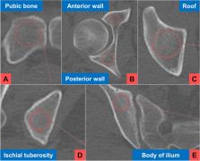
          This study enrolled young patients with high-energy trauma (aged 20–44 years; young group) and older patients with low-energy trauma (aged 65–89 years; older group). Patients without pelvic computed tomography (CT) scans, those with pelvic bone implants, and those who died were excluded. The HU values on the contralateral (non-fractured) side of the pelvis were measured on CT scans. The CT data were divided into 7 areas: the pubic bone, the anterior and posterior walls and roof of the acetabulum, the ischial tuberosity, the body of the ilium, and the third lumbar vertebra. The HU values in each area were compared between the young and older groups.

          Results

          Sixty-one young patients and 154 older patients were included in the study. The highest HU value was in the roof of the acetabulum regardless of age and sex. HU values were significantly higher in the ischial tuberosity and body of the ilium and lower in the pubic bone and anterior wall. The HU values in all pelvic areas were significantly lower in the older group than in the young group, especially in the anterior area.

          Conclusions

          HU values in the 6 pelvic areas were not uniform and were strongly related to load distribution. The HU distribution and age-related differences could explain the characteristic causes and patterns of acetabular fractures in the older and may help in surgical treatment.

          Related collections

          Most cited references38

          • Record: found
          • Abstract: found
          • Article: not found

          Hounsfield units for assessing bone mineral density and strength: a tool for osteoporosis management.

          Measurements obtained from clinical computed tomography examinations may yield information leading to the diagnosis of decreased bone mineral density, without added expense to the patient. The purpose of the present study was to determine if Hounsfield units, a standardized computed tomography attenuation coefficient, correlate with bone mineral density and compressive strength.
            Bookmark
            • Record: found
            • Abstract: found
            • Article: not found

            Fractures of the acetabulum in patients aged 60 years and older: an epidemiological and radiological study.

            Using a prospective database of 1309 displaced acetabular fractures gathered between 1980 and 2007, we calculated the annual mean age and annual incidence of elderly patients > 60 years of age presenting with these injuries. We compared the clinical details and patterns of fracture between patients > 60 years of age (study group) with those 60 years of age and the remaining 1074 were < 60 years. The incidence of elderly patients with acetabular fractures increased by 2.4-fold between the first half of the study period and the second half (10% (62) vs 24% (174), p < 0.001). Fractures characterised by displacement of the anterior column were significantly more common in the elderly compared with the younger patients (64% (150) vs 43% (462), respectively, p < 0.001). Common radiological features of the fractures in the study group included a separate quadrilateral-plate component (50.8% (58)) and roof impaction (40% (46)) in the anterior fractures, and comminution (44% (30)) and marginal impaction (38% (26)) in posterior-wall fractures. The proportion of elderly patients presenting with acetabular fractures increased during the 27-year period. The older patients had a different distribution of fracture pattern than the younger patients, and often had radiological features which have been shown in other studies to be predictive of a poor outcome.
              Bookmark
              • Record: found
              • Abstract: found
              • Article: not found

              Predictive validity of preoperative CT scans and the risk of pedicle screw loosening in spinal surgery.

              Pedicle screw fixation is the standard technique for the stabilization of the spine, a clinically relevant complication of which is screw loosening. This retrospective study investigates whether preoperative CT scanning can offer a predictor of screw loosening.
                Bookmark

                Author and article information

                Contributors
                n.inagaki@jikei.ac.jp
                tanaka.takaaki.vh@mail.hosp.go.jp
                judk@jikei.ac.jp
                akiyamashoshi@gmail.com
                ms02-matsuoka@jikei.ac.jp
                xlink67@gol.com
                Journal
                BMC Musculoskelet Disord
                BMC Musculoskelet Disord
                BMC Musculoskeletal Disorders
                BioMed Central (London )
                1471-2474
                29 March 2022
                29 March 2022
                2022
                : 23
                : 305
                Affiliations
                [1 ]GRID grid.411898.d, ISNI 0000 0001 0661 2073, Department of Orthopedic Surgery, , The Jikei University School of Medicine, ; Tokyo, Japan
                [2 ]Department of Orthopedic Surgery, NHO Utsunomiya National Hospital, Tochigi, Japan
                Article
                5263
                10.1186/s12891-022-05263-3
                8966165
                35351073
                d0404855-3e80-416d-9497-198de7b66ffd
                © The Author(s) 2022

                Open AccessThis article is licensed under a Creative Commons Attribution 4.0 International License, which permits use, sharing, adaptation, distribution and reproduction in any medium or format, as long as you give appropriate credit to the original author(s) and the source, provide a link to the Creative Commons licence, and indicate if changes were made. The images or other third party material in this article are included in the article's Creative Commons licence, unless indicated otherwise in a credit line to the material. If material is not included in the article's Creative Commons licence and your intended use is not permitted by statutory regulation or exceeds the permitted use, you will need to obtain permission directly from the copyright holder. To view a copy of this licence, visit http://creativecommons.org/licenses/by/4.0/. The Creative Commons Public Domain Dedication waiver ( http://creativecommons.org/publicdomain/zero/1.0/) applies to the data made available in this article, unless otherwise stated in a credit line to the data.

                History
                : 1 May 2021
                : 24 March 2022
                Categories
                Research
                Custom metadata
                © The Author(s) 2022

                Orthopedics
                pelvic fracture,bone mineral density,hounsfield unit,fragility fracture
                Orthopedics
                pelvic fracture, bone mineral density, hounsfield unit, fragility fracture

                Comments

                Comment on this article