0
views
0
recommends
+1 Recommend
0 collections
    0
    shares
      • Record: found
      • Abstract: found
      • Article: found
      Is Open Access

      Value of Hounsfield units measured by chest computed tomography for assessing bone density in the thoracolumbar segment of the thoracic spine

      research-article

      Read this article at

      Bookmark
          There is no author summary for this article yet. Authors can add summaries to their articles on ScienceOpen to make them more accessible to a non-specialist audience.

          Abstract

          Study Design

          A retrospective study.

          Purpose

          To investigate the correlation between Hounsfield unit (HU) values measured by chest computed tomography (CT) and dual-energy X-ray absorptiometry (DXA) T-scores. HU-based thoracolumbar (T11 and T12) cutoff thresholds were calculated for a cohort of Chinese patients.

          Overview of Literature

          For patients with osteoporosis, the incidence of fractures in the thoracolumbar segment is significantly higher than that in other sites. However, most current clinical studies have focused on L1.

          Methods

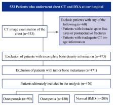
          This retrospective study analyzed patients who underwent chest CT and DXA at our hospital between August 2021 and August 2022. Thoracic thoracolumbar segment HU values, lumbar T-scores, and hip T-scores were computed for comparison, and thoracic thoracolumbar segment HU thresholds suggestive of potential bone density abnormalities were established using receiver operating characteristic curves.

          Results

          In total, 470 patients (72.4% women; mean age, 65.5±12.3 years) were included in this study. DXA revealed that of the 470 patients, 90 (19%) had osteoporosis, 180 (38%) had reduced osteopenia, and 200 (43%) had normal bone mineral density (BMD). To differentiate osteoporosis from osteopenia, the HU threshold was established as 105.1 (sensitivity, 54.4%; specificity, 72.2%) for T11 and 85.7 (sensitivity, 69.4%; specificity, 61.1%) for T12. To differentiate between osteopenia and normal BMD, the HU threshold was 146.7 for T11 (sensitivity, 57.5%; specificity, 84.4%) and 135.7 for T12 (sensitivity, 59.5%; specificity, 80%).

          Conclusions

          This study supports the significance of HU values from chest CT for BMD assessment. Chest CT provides a new method for clinical opportunistic screening of osteoporosis. When the T11 HU is >146.7 or the T12 HU is >135.7, additional osteoporosis testing is not needed unless a vertebral fracture is detected. If the T11 HU is <105.1 or the T12 HU is <85.7, further DXA testing is strongly advised. In addition, vertebral HU values that fall faster than those of the T11 and L1 vertebrae may explain the high incidence of T12 vertebral fractures.

          Related collections

          Most cited references34

          • Record: found
          • Abstract: found
          • Article: found
          Is Open Access

          Prevalence of osteoporosis in China: a meta-analysis and systematic review

          Background We conducted a systematic review and meta-analysis to obtain a reliable estimation of the prevalence of osteoporosis in China and to characterize its epidemiology. Methods We identified relevant studies via a search of literature published from 2003 to October 2015 in the PubMed, Web of Science, China National Knowledge Infrastructure, Wanfang and Weipu databases. Both Chinese and WHO criteria were considered acceptable for the diagnosis of osteoporosis. Prevalence estimates were obtained using random effects models. Meta-regression analysis was used to explore the sources of heterogeneity, and publication bias was evaluated by visually inspecting funnel plots. Results Overall, 69 articles were included in this study. An obvious increase in the prevalence of osteoporosis was identified over the past 12 years (prevalence of 14.94 % before 2008 and 27.96 % during the period spanning 2012–2015). The prevalence of osteoporosis was higher in females than in males (25.41 % vs. 15.33 %) and increased with age. Osteoporosis prevalence was higher in rural than in urban areas (20.87 % vs. 23.92 %) and higher in southern than in northern areas (23.17 % vs. 20.13 %). At present, the pooled prevalence of osteoporosis in people aged 50 years and older was more than twice the pooled prevalence identified in 2006 (34.65 % vs. 15.7 %). The application of different diagnostic criteria could have an impact on prevalence estimation (19.7 % vs. 29.3 %). Meta-regression suggested that study setting also influenced the estimation of point prevalence (P = 0.022). Conclusions The prevalence of osteoporosis in China has increased over the past 12 years, affecting more than one-third of people aged 50 years and older. The prevalence of osteoporosis increased with age and was higher in females than in males. Prevention and control measures have become all the more important given the increase in osteoporosis prevalence, and three-step prevention programmes should be implemented. Electronic supplementary material The online version of this article (doi:10.1186/s12889-016-3712-7) contains supplementary material, which is available to authorized users.
            Bookmark
            • Record: found
            • Abstract: found
            • Article: not found

            Bone microarchitecture assessed by TBS predicts osteoporotic fractures independent of bone density: the Manitoba study.

            The measurement of BMD by dual-energy X-ray absorptiometry (DXA) is the "gold standard" for diagnosing osteoporosis but does not directly reflect deterioration in bone microarchitecture. The trabecular bone score (TBS), a novel gray-level texture measurement that can be extracted from DXA images, correlates with 3D parameters of bone microarchitecture. Our aim was to evaluate the ability of lumbar spine TBS to predict future clinical osteoporotic fractures. A total of 29,407 women 50 years of age or older at the time of baseline hip and spine DXA were identified from a database containing all clinical results for the Province of Manitoba, Canada. Health service records were assessed for the incidence of nontraumatic osteoporotic fracture codes subsequent to BMD testing (mean follow-up 4.7 years). Lumbar spine TBS was derived for each spine DXA examination blinded to clinical parameters and outcomes. Osteoporotic fractures were identified in 1668 (5.7%) women, including 439 (1.5%) spine and 293 (1.0%) hip fractures. Significantly lower spine TBS and BMD were identified in women with major osteoporotic, spine, and hip fractures (all p < 0.0001). Spine TBS and BMD predicted fractures equally well, and the combination was superior to either measurement alone (p < 0.001). Spine TBS predicts osteoporotic fractures and provides information that is independent of spine and hip BMD. Combining the TBS trabecular texture index with BMD incrementally improves fracture prediction in postmenopausal women. Copyright © 2011 American Society for Bone and Mineral Research.
              Bookmark
              • Record: found
              • Abstract: found
              • Article: not found

              Prevention and management of osteoporosis.

              (2002)
              Bone is hard tissue that is in a constant state of flux, being built up by bone-forming cells called osteoblasts while also being broken down or resorbed by cells known as osteoclasts. During childhood and adolescence, bone formation is dominant; bone length and girth increase with age, ending at early adulthood when peak bone mass is attained. Males generally exhibit a longer growth period, resulting in bones of greater size and overall strength. In males after the age of 20, bone resorbtion becomes predominant, and bone mineral content declines about 4% per decade. Females tend to maintain peak mineral content until menopause, after which time it declines about 15% per decade. Osteoporosis is a disease characterized by low bone mass and structural deterioration of bone tissue, leading to bone fragility and an increased susceptibility to fractures, especially of the hip, spine, and wrist. Osteoporosis occurs primarily as a result of normal ageing, but can arise as a result of impaired development of peak bone mass (e.g. due to delayed puberty or undernutrition) or excessive bone loss during adulthood (e.g. due to estrogen deficiency in women, undernutrition, or corticosteroid use). Osteoporosis-induced fractures cause a great burden to society. Hip fractures are the most serious, as they nearly always result in hospitalization, are fatal about 20% of the time, and produce permanent disability about half the time. Fracture rates increase rapidly with age and the lifetime risk of fracture in 50 year-old women is about 40%, similar to that for coronary heart disease. In 1990, there were 1.7 million hip fractures alone worldwide; with changes in population demographics, this figure is expected to rise to 6 million by 2050. To help describe the nature and consequences of osteoporosis, as well as strategies for its prevention and management, a WHO Scientific Group meeting of international experts was held in Geneva, which resulted in this technical report. This monograph describes in detail normal bone development and the causes and risk factors for developing osteoporosis. The burden of osteoporosis is characterized in terms of mortality, morbidity, and economic costs. Methods for its prevention and treatment are discussed in detail for both pharmacological and non-pharmacological approaches. For each approach, the strength of the scientific evidence is presented. The report also provides cost-analysis information for potential interventions, and discusses important aspects of developing national policies to deal with osteoporosis. Recommendations are made to the general population, care providers, health administrators, and researchers. Lastly, national organizations and support groups are listed by country.
                Bookmark

                Author and article information

                Journal
                Asian Spine J
                Asian Spine J
                Asian Spine Journal
                Korean Society of Spine Surgery
                1976-1902
                1976-7846
                June 2024
                25 June 2024
                : 18
                : 3
                : 336-345
                Affiliations
                [1 ]Department of Spine Surgery, Affiliated Hospital of Integrated Traditional Chinese and Western Medicine, Nanjing University of Chinese Medicine, Nanjing, P.R. China
                [2 ]Department of Imaging, Affiliated Hospital of Integrated Traditional Chinese and Western Medicine, Nanjing University of Chinese Medicine, Nanjing, P.R. China
                [3 ]Department of Orthopaedics, Traditional Chinese Medicine Hospital of Ili Kazak Autonomous Prefecture, Yining, China
                Author notes
                Corresponding author: Zhipeng Xi, Department of Spine Surgery, Affiliated Hospital of Integrated Traditional Chinese and Western Medicine, Nanjing University of Chinese Medicine, 100th Shizi Street, Nanjing, 210028, Jiangsu Province, P.R. China, Tel: +86-13675154279, Fax: +86-02585502829, E-mail: xizhipeng1985@ 123456163.com
                Co-corresponding author: Yubo Wei, Department of Imaging, Affiliated Hospital of Integrated Traditional Chinese and Western Medicine, Nanjing University of Chinese Medicine, 100th Shizi Street, Nanjing, 210028, Jiangsu Province, P.R. China, Tel: +86-13951667096, Fax: +86-02585502829, E-mail: 13951667096@ 123456163.com
                [*]

                These authors contributed equally to this work as corresponding authors.

                Author information
                https://orcid.org/0009-0004-5683-2065
                https://orcid.org/0009-0007-3597-4832
                https://orcid.org/0000-0002-7605-8634
                https://orcid.org/0000-0001-7124-1029
                https://orcid.org/0009-0002-0540-3098
                https://orcid.org/0009-0000-3046-8681
                https://orcid.org/0000-0003-2021-4842
                Article
                asj-2023-0438
                10.31616/asj.2023.0438
                11222884
                38917853
                2c093598-e12d-4e3c-b441-60199d291f87
                Copyright © 2024 by Korean Society of Spine Surgery

                This is an Open Access article distributed under the terms of the Creative Commons Attribution Non-Commercial License ( http://creativecommons.org/licenses/by-nc/4.0/) which permits unrestricted non-commercial use, distribution, and reproduction in any medium, provided the original work is properly cited.

                History
                : 2 January 2024
                : 31 January 2024
                : 6 February 2024
                Categories
                Clinical Study

                Orthopedics
                bone density,chest computed tomography,osteoporosis,hounsfield unit,thoracolumbar segment of the thoracic spine

                Comments

                Comment on this article