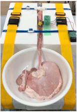
Endoscopic submucosal dissection (ESD) is an established endoscopic resection method in Asian countries, which is increasingly practiced in Europe and by early adopters in the United States for removal of early cancers and large lesions from the luminal gastrointestinal tract. The intent of this expert review is to provide an update regarding the clinical practice of ESD with a particular focus on its use in the United States. This review is framed around the 16 best practice advice points agreed upon by the authors, which reflect landmark and recent published articles in this field. This expert review also reflects our experience as advanced endoscopists with extensive experience in performing and teaching others to perform ESD in the United States. Best Practice Advice 1: Endoscopic submucosal dissection should be recognized as a mature endoscopic technique that enables complete removal of lesions that are too large for en bloc endoscopic mucosal resection or are at increased risk of containing cancer. Best Practice Advice 2: The safety and feasibility of endoscopic submucosal dissection for early gastric cancer is well established. The absolute indications for curative endoscopic resection include moderately and well-differentiated, nonulcerated, mucosal lesions that are ≤2 cm in size. Best Practice Advice 3: Other relative (expanded) indications for gastric endoscopic submucosal dissection include moderately and well-differentiated superficial cancers that are >2 cm, lesions ≤3 cm with ulceration or that contain early submucosal invasion, and poorly differentiated superficial cancers ≤2 cm in size. The risk of lymph node metastasis when endoscopic submucosal dissection is performed for these indications is higher than when it is performed for absolute indications but remains acceptably low. Best Practice Advice 4: Endoscopic submucosal dissection may be considered in selected patients with Barrett's esophagus with the following features: large or bulky area of nodularity, lesions with a high likelihood of superficial submucosal invasion, recurrent dysplasia, endoscopic mucosal resection specimen showing invasive carcinoma with positive margins, equivocal preprocedural histology, and intramucosal carcinoma. Best Practice Advice 5: Endoscopic submucosal dissection is the primary modality for treatment of squamous cell dysplasia and cancer confined to the superficial esophageal mucosa. Any degree of submucosal invasion caries an increased risk of lymph node metastasis and alternative/additional therapy should be considered. Best Practice Advice 6: Duodenal endoscopic submucosal dissection is associated with an increased risk of intraprocedural perforation and delayed adverse events. Duodenal endoscopic submucosal dissection should be limited to endoscopists with extensive experience in performing endoscopic submucosal dissection in other locations. It is strongly suggested that endoscopists in the United States refrain from performing duodenal endoscopic submucosal dissection during the early phase of their endoscopic submucosal dissection practice. Best Practice Advice 7: All colorectal lesions should be evaluated for suitability for endoscopic resection. Accumulating evidence has shown that the majority of colorectal neoplasms without signs of deep submucosal invasion or advanced cancer can be treated by advanced endoscopic resection techniques. Best Practice Advice 8: Colorectal neoplasms containing dysplasia confined to the mucosa have no risk for lymph node metastasis and endoscopic resection should be considered as the criterion standard. Best Practice Advice 9: Large (>2 cm) colorectal lesions frequently (>43%) require piecemeal removal when endoscopic mucosal resection is used, which is associated with increased (up to 20%) rates of recurrent neoplasia. Endoscopic submucosal dissection enables higher rates of en bloc resection and lower recurrence rates for these lesions. Patients with large complex colorectal polyps should be referred to a high-volume, specialized center for endoscopic removal by endoscopic mucosal resection or endoscopic submucosal dissection. Best Practice Advice 10: Endoscopic resection for colorectal lesions offers significant cost benefit compared with surgery, and case-based endoscopic submucosal dissection selection for high-risk lesions could offer cost savings. Best Practice Advice 11: Endoscopists in the United States embarking on performing endoscopic submucosal dissection should be familiar with currently available endoscopic tissue closure devices. Both clip closure and endoscopic suturing techniques have been shown to be effective in managing intraprocedural perforation. Complete closure of a post-endoscopic submucosal dissection site may be considered in certain circumstances based on patient factors, procedural factors, and the location of the lesion. Best Practice Advice 12: Careful coagulation of exposed blood vessels in the resection site may reduce the risk of delayed bleeding after endoscopic submucosal dissection. The use of low-voltage coagulation current is recommended for this technique. Best Practice Advice 13: Endoscopists should affix the endoscopic submucosal dissection specimen to a flat surface (eg, pin the specimen to cork board) and immerse it in formalin. An expert gastrointestinal pathologist should evaluate the specimen for margin involvement, degree of differentiation, presence or absence of lymphovascular invasion, depth of submucosal invasion (if present), and tumor budding. Best Practice Advice 14: Acquiring high-level competency in endoscopic submucosal dissection is achievable in the United States. Alternative educational models should be used in the United States because of the limited number of experts and the differing prevalence of gastrointestinal luminal diseases as compared with Asia. Best Practice Advice 15: The endoscopic submucosal dissection educational model most suited for the current environment in the United States is a stepwise approach consisting of didactic self-study, attending training courses with increasing levels of complexity, self-practice on animal models, and observation of live cases performed by experts. Endoscopists should perform their initial endoscopic submucosal dissections on patients with lesions that have well-established indications for endoscopic submucosal dissection and are of the lowest technical complexity. Best Practice Advice 16: Endoscopists in the United States who perform endoluminal resection should educate referring physicians to avoid practices that may induce submucosal fibrosis hampering future endoscopic mucosal resection or endoscopic submucosal dissection. These practices include tattooing in close proximity to or beneath a lesion for marking and partial snare resection of a portion of a lesion for histopathology.