8
views
0
recommends
+1 Recommend
0 collections
    0
    shares
      • Record: found
      • Abstract: found
      • Article: found
      Is Open Access

      Copper Ions and Parkinson’s Disease: Why Is Homeostasis So Relevant?

      review-article
      * , *
      Biomolecules
      MDPI
      copper, dopamine, Parkinson’s disease, α-synuclein, SOD1

      Read this article at

      Bookmark
          There is no author summary for this article yet. Authors can add summaries to their articles on ScienceOpen to make them more accessible to a non-specialist audience.

          Abstract

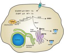
          The involvement of copper in numerous physiological processes makes this metal ion essential for human life. Alterations in copper homeostasis might have deleterious consequences, and several neurodegenerative disorders, including Parkinson’s disease (PD), have been associated with impaired copper levels. In the present review, we describe the molecular mechanisms through which copper can exert its toxicity, by considering how it can interfere with other cellular processes known to play a role in PD, such as dopamine metabolism, oxidative stress, and α-synuclein aggregation. The recent experimental evidence that associates copper deficiency and the formation of superoxide dismutase 1 (SOD1) aggregates with the progression of PD is also discussed together with its therapeutic implication. Overall, the recent discoveries described in this review show how either copper deficiency or excessive levels can promote detrimental effects, highlighting the importance of preserving copper homeostasis and opening unexplored therapeutic avenues in the definition of novel disease-modifying drugs.

          Related collections

          Most cited references68

          • Record: found
          • Abstract: found
          • Article: not found

          Targeting chelatable iron as a therapeutic modality in Parkinson's disease.

          The pathophysiological role of iron in Parkinson's disease (PD) was assessed by a chelation strategy aimed at reducing oxidative damage associated with regional iron deposition without affecting circulating metals. Translational cell and animal models provided concept proofs and a delayed-start (DS) treatment paradigm, the basis for preliminary clinical assessments. For translational studies, we assessed the effect of oxidative insults in mice systemically prechelated with deferiprone (DFP) by following motor functions, striatal dopamine (HPLC and MRI-PET), and brain iron deposition (relaxation-R2*-MRI) aided by spectroscopic measurements of neuronal labile iron (with fluorescence-sensitive iron sensors) and oxidative damage by markers of protein, lipid, and DNA modification. DFP significantly reduced labile iron and biological damage in oxidation-stressed cells and animals, improving motor functions while raising striatal dopamine. For a pilot, double-blind, placebo-controlled randomized clinical trial, early-stage Parkinson's patients on stabilized dopamine regimens enrolled in a 12-month single-center study with DFP (30 mg/kg/day). Based on a 6-month DS paradigm, early-start patients (n=19) compared to DS patients (n=18) (37/40 completed) responded significantly earlier and sustainably to treatment in both substantia nigra iron deposits (R2* MRI) and Unified Parkinson's Disease Rating Scale motor indicators of disease progression (p<0.03 and p<0.04, respectively). Apart from three rapidly resolved neutropenia cases, safety was maintained throughout the trial. A moderate iron chelation regimen that avoids changes in systemic iron levels may constitute a novel therapeutic modality for PD. The therapeutic features of a chelation modality established in translational models and in pilot clinical trials warrant comprehensive evaluation of symptomatic and/or disease-modifying potential of chelation in PD.
            Bookmark
            • Record: found
            • Abstract: found
            • Article: not found

            Increased nigral iron content and alterations in other metal ions occurring in brain in Parkinson's disease.

            Levels of iron, copper, zinc, manganese, and lead were measured by inductively coupled plasma spectroscopy in parkinsonian and age-matched control brain tissue. There was 31-35% increase in the total iron content of the parkinsonian substantia nigra when compared to control tissue. In contrast, in the globus pallidus total iron levels were decreased by 29% in Parkinson's disease. There was no change in the total iron levels in any other region of the parkinsonian brain. Total copper levels were reduced by 34-45% in the substantia nigra in Parkinson's disease; no difference was found in the other brain areas examined. Zinc levels were increased in substantia nigra in Parkinson's disease by 50-54%, and the zinc content of the caudate nucleus and lateral putamen was also raised by 18-35%. Levels of manganese and lead were unchanged in all areas of the parkinsonian brain studied when compared to control brains, except for a small decrease (20%) in manganese content of the medial putamen. Increased levels of total iron in the substantia nigra may cause the excessive formation of toxic oxygen radicals, leading to dopamine cell death.
              Bookmark
              • Record: found
              • Abstract: found
              • Article: not found

              Transition metals, ferritin, glutathione, and ascorbic acid in parkinsonian brains.

              The regional distributions of iron, copper, zinc, magnesium, and calcium in parkinsonian brains were compared with those of matched controls. In mild Parkinson's disease (PD), there were no significant differences in the content of total iron between the two groups, whereas there was a significant increase in total iron and iron (III) in substantia nigra of severely affected patients. Although marked regional distributions of iron, magnesium, and calcium were present, there were no changes in magnesium, calcium, and copper in various brain areas of PD. The most notable finding was a shift in the iron (II)/iron (III) ratio in favor of iron (III) in substantia nigra and a significant increase in the iron (III)-binding, protein, ferritin. A significantly lower glutathione content was present in pooled samples of putamen, globus pallidus, substantia nigra, nucleus basalis of Meynert, amygdaloid nucleus, and frontal cortex of PD brains with severe damage to substantia nigra, whereas no significant changes were observed in clinicopathologically mild forms of PD. In all these regions, except the amygdaloid nucleus, ascorbic acid was not decreased. Reduced glutathione and the shift of the iron (II)/iron (III) ratio in favor of iron (III) suggest that these changes might contribute to pathophysiological processes underlying PD.
                Bookmark

                Author and article information

                Journal
                Biomolecules
                Biomolecules
                biomolecules
                Biomolecules
                MDPI
                2218-273X
                29 January 2020
                February 2020
                : 10
                : 2
                : 195
                Affiliations
                Molecular Physiology and Biophysics Unit, Department of Biology, Università di Padova, 35131 Padova, Italy
                Author notes
                Author information
                https://orcid.org/0000-0002-3968-8418
                https://orcid.org/0000-0001-7927-9208
                Article
                biomolecules-10-00195
                10.3390/biom10020195
                7072482
                32013126
                ccaea366-57c1-43ce-80a5-e07335d4babe
                © 2020 by the authors.

                Licensee MDPI, Basel, Switzerland. This article is an open access article distributed under the terms and conditions of the Creative Commons Attribution (CC BY) license ( http://creativecommons.org/licenses/by/4.0/).

                History
                : 27 December 2019
                : 28 January 2020
                Categories
                Review

                copper,dopamine,parkinson’s disease,α-synuclein,sod1
                copper, dopamine, parkinson’s disease, α-synuclein, sod1

                Comments

                Comment on this article