23
views
0
recommends
+1 Recommend
0 collections
    0
    shares
      • Record: found
      • Abstract: found
      • Article: found
      Is Open Access

      Neural stem cell-derived small extracellular vesicles attenuate apoptosis and neuroinflammation after traumatic spinal cord injury by activating autophagy

      research-article

      Read this article at

      Bookmark
          There is no author summary for this article yet. Authors can add summaries to their articles on ScienceOpen to make them more accessible to a non-specialist audience.

          Abstract

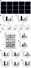
          Spinal cord injury (SCI) can cause severe irreversible motor dysfunction and even death. Neural stem cell (NSC) transplantation can promote functional recovery after acute SCI in experimental animals, but numerous issues, including low-transplanted cell survival rate, cell de-differentiation, and tumor formation need to be resolved before routine clinical application is feasible. Recent studies have shown that transplanted stem cells facilitate regeneration through release of paracrine factors. Small extracellular vesicles (sEVs), the smallest known membrane-bound nanovesicles, are involved in complex intercellular communication systems and are an important vehicle for paracrine delivery of therapeutic agents. However, the application of NSC-derived small extracellular vesicles (NSC-sEVs) to SCI treatment has not been reported. We demonstrate that NSC-sEVs can significantly reduce the extent of SCI, improve functional recovery, and reduce neuronal apoptosis, microglia activation, and neuroinflammation in rats. Furthermore, our study suggests that NSC-sEVs can regulate apoptosis and inflammatory processes by inducing autophagy. In brief, NSC-sEVs increased the expression of the autophagy marker proteins LC3B and beclin-1, and promoted autophagosome formation. Following NSC-sEV infusion, the SCI area was significantly reduced, and the expression levels of the proapoptotic protein Bax, the apoptosis effector cleaved caspase-3, and the pro-inflammatory cytokines TNF-α, IL-1β, and IL-6 were significantly reduced, whereas the expression level of the anti-apoptotic protein Bcl-2 was upregulated. In the presence of the autophagy inhibitor 3MA, however, these inhibitory effects of NSC-sEVs on apoptosis and neuroinflammation were significantly reversed. Our results show for the first time that NSC-sEV treatment has the potential to reduce neuronal apoptosis, inhibit neuroinflammation, and promote functional recovery in SCI model rats at an early stage by promoting autophagy.

          Related collections

          Most cited references48

          • Record: found
          • Abstract: found
          • Article: not found

          Hypothalamic stem cells control aging speed partly through exosomal miRNAs

          SUMMARY Hypothalamic control of aging was recently proposed, but the responsible mechanisms still remain unclear. Here, following the observation that aging of mice started with a substantial loss of hypothalamic stem/progenitor cells that co-express Sox2 and Bmi1, we developed several mouse models with ablation of these hypothalamic cells, each of them consistently displaying an acceleration in aging-like physiological changes or shortening in lifespan. Conversely, aging retardation and lifespan extension were achieved in mid-aged mice when locally implanted with healthy hypothalamic stem/progenitor cells that were genetically engineered to survive from aging-related hypothalamic inflammatory microenvironment. Mechanistically, hypothalamic stem/progenitor cells greatly contributed to exosomal miRNAs in the cerebrospinal fluid which declined over aging, while central treatment with healthy hypothalamic stem/progenitor cells-secreted exosomes led to slowdown of aging. In conclusion, aging speed is controlled significantly by hypothalamic stem cells partially through release of exosomal miRNAs.
            Bookmark
            • Record: found
            • Abstract: found
            • Article: not found

            Haematopoietic stem cells adopt mature haematopoietic fates in ischaemic myocardium.

            Under conditions of tissue injury, myocardial replication and regeneration have been reported. A growing number of investigators have implicated adult bone marrow (BM) in this process, suggesting that marrow serves as a reservoir for cardiac precursor cells. It remains unclear which BM cell(s) can contribute to myocardium, and whether they do so by transdifferentiation or cell fusion. Here, we studied the ability of c-kit-enriched BM cells, Lin- c-kit+ BM cells and c-kit+ Thy1.1(lo) Lin- Sca-1+ long-term reconstituting haematopoietic stem cells to regenerate myocardium in an infarct model. Cells were isolated from transgenic mice expressing green fluorescent protein (GFP) and injected directly into ischaemic myocardium of wild-type mice. Abundant GFP+ cells were detected in the myocardium after 10 days, but by 30 days, few cells were detectable. These GFP+ cells did not express cardiac tissue-specific markers, but rather, most of them expressed the haematopoietic marker CD45 and myeloid marker Gr-1. We also studied the role of circulating cells in the repair of ischaemic myocardium using GFP+-GFP- parabiotic mice. Again, we found no evidence of myocardial regeneration from blood-borne partner-derived cells. Our data suggest that even in the microenvironment of the injured heart, c-kit-enriched BM cells, Lin- c-kit+ BM cells and c-kit+ Thy1.1(lo) Lin- Sca-1+ long-term reconstituting haematopoietic stem cells adopt only traditional haematopoietic fates.
              Bookmark
              • Record: found
              • Abstract: found
              • Article: not found

              Spontaneous human adult stem cell transformation.

              Human adult stem cells are being evaluated widely for various therapeutic approaches. Several recent clinical trials have reported their safety, showing them to be highly resistant to transformation. The clear similarities between stem cell and cancer stem cell genetic programs are nonetheless the basis of a recent proposal that some cancer stem cells could derive from human adult stem cells. Here we show that although they can be managed safely during the standard ex vivo expansion period (6-8 weeks), human mesenchymal stem cells can undergo spontaneous transformation following long-term in vitro culture (4-5 months). This is the first report of spontaneous transformation of human adult stem cells, supporting the hypothesis of cancer stem cell origin. Our findings indicate the importance of biosafety studies of mesenchymal stem cell biology to efficiently exploit their full clinical therapeutic potential.
                Bookmark

                Author and article information

                Contributors
                +8613951661018 , caiwhspine@sina.com
                Journal
                Cell Death Dis
                Cell Death Dis
                Cell Death & Disease
                Nature Publishing Group UK (London )
                2041-4889
                18 April 2019
                18 April 2019
                May 2019
                : 10
                : 5
                : 340
                Affiliations
                [1 ]ISNI 0000 0004 1799 0784, GRID grid.412676.0, Department of Orthopaedics, , First Affiliated Hospital of Nanjing Medical University, ; Nanjing, 210029 Jiangsu China
                [2 ]ISNI 0000 0004 1770 1022, GRID grid.412901.f, Department of Orthopaedics, , West China Hospital Sichuan University, ; Chengdu, 610000 Sichuan China
                Article
                1571
                10.1038/s41419-019-1571-8
                6472377
                31000697
                cc3ad641-f79a-4f6c-b393-567915be6b0f
                © The Author(s) 2019

                Open Access This article is licensed under a Creative Commons Attribution 4.0 International License, which permits use, sharing, adaptation, distribution and reproduction in any medium or format, as long as you give appropriate credit to the original author(s) and the source, provide a link to the Creative Commons license, and indicate if changes were made. The images or other third party material in this article are included in the article’s Creative Commons license, unless indicated otherwise in a credit line to the material. If material is not included in the article’s Creative Commons license and your intended use is not permitted by statutory regulation or exceeds the permitted use, you will need to obtain permission directly from the copyright holder. To view a copy of this license, visit http://creativecommons.org/licenses/by/4.0/.

                History
                : 1 February 2019
                : 15 March 2019
                : 3 April 2019
                Categories
                Article
                Custom metadata
                © The Author(s) 2019

                Cell biology
                cell death in the nervous system,neural stem cells
                Cell biology
                cell death in the nervous system, neural stem cells

                Comments

                Comment on this article