7
views
0
recommends
+1 Recommend
0 collections
    0
    shares
      • Record: found
      • Abstract: found
      • Article: found
      Is Open Access

      IGF-1C domain–modified hydrogel enhanced the efficacy of stem cells in the treatment of AMI

      research-article

      Read this article at

      Bookmark
          There is no author summary for this article yet. Authors can add summaries to their articles on ScienceOpen to make them more accessible to a non-specialist audience.

          Abstract

          Background

          Due to the low survival rate of cell transplantation, stem cell has not been widely used in clinical treatment of acute myocardial infarction (AMI). In this study, we immobilized the C domain peptide of insulin-like growth factor-1 on chitosan (CS-IGF-1C) to obtain bioactive hydrogel. The purpose was to investigate whether CS-IGF-1C hydrogel incorporated with human placenta–derived mesenchymal stem cells (hP-MSCs) can boost the survival of hP-MSCs and enhance their therapeutic effects.

          Methods

          hP-MSCs, which continuously expressed green fluorescent protein (GFP) and firefly luciferase (Fluc), were transplanted with CS-IGF-1C hydrogel into a mouse myocardial infarction model. Cell survival was detected by bioluminescence imaging (BLI), and cardiac function was measured by echocardiogram. Real-time PCR and histological analysis were used to explore the therapeutic mechanism of CS-IGF-1C hydrogel.

          Results

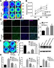
          CS-IGF-1C hydrogel could induce the proliferation of hP-MSCs and exert anti-apoptotic effects in vitro. The Calcine-AM/PI staining results showed that hP-MSCs seeded on CS-IGF-1C hydrogel could protect neonatal mouse ventricular cardiomyocytes (NMVCs) against oxidative stress. It was observed by BLI that CS-IGF-1C hydrogel injected into ischemic myocardium could improve the survival rate of hP-MSCs. Histology analysis indicated that co-transplantation of the CS-IGF-1C hydrogel and hP-MSCs could increase angiogenesis, reduce collagen deposition, ameliorate left ventricular expanded, and further promote the recovery of cardiac function. Besides, we found that the inflammatory response was inhibited and the expression of apoptosis-related genes was downregulated by CS-IGF-1C hydrogel.

          Conclusions

          CS-IGF-1C hydrogel provides a conducive microenvironment for cells and significantly boosts the survival of hP-MSCs in mouse myocardial infarction model, which suggest that it may be a potential candidate for prolonging the therapeutic effect of hP-MSCs during AMI.

          Related collections

          Most cited references25

          • Record: found
          • Abstract: found
          • Article: not found

          A novel and efficient model of coronary artery ligation and myocardial infarction in the mouse.

          coronary artery ligation to induce myocardial infarction (MI) in mice is typically performed by an invasive and time-consuming approach that requires ventilation and chest opening (classic method), often resulting in extensive tissue damage and high mortality. We developed a novel and rapid surgical method to induce MI that does not require ventilation. the purpose of this study was to develop and comprehensively describe this method and directly compare it to the classic method. male C57/B6 mice were grouped into 4 groups: new method MI (MI-N) or sham (S-N) and classic method MI (MI-C) or sham (S-C). In the new method, heart was manually exposed without intubation through a small incision and MI was induced. In the classic method, MI was induced through a ventilated thoracotomy. Similar groups were used in an ischemia/reperfusion injury model. This novel MI procedure is rapid, with an average procedure time of 1.22 ± 0.05 minutes, whereas the classic method requires 23.2 ± 0.6 minutes per procedure. Surgical mortality was 3% in MI-N and 15.9% in MI-C. The rate of arrhythmia was significantly lower in MI-N. The postsurgical levels of tumor necrosis factor-α and myeloperoxidase were lower in new method, indicating less inflammation. Overall, 28-day post-MI survival rate was 68% with MI-N and 48% with MI-C. Importantly, there was no difference in infarct size or post-MI cardiac function between the methods. this new rapid method of MI in mice represents a more efficient and less damaging model of myocardial ischemic injury compared with the classic method.
            Bookmark
            • Record: found
            • Abstract: found
            • Article: not found

            IGF-1-overexpressing mesenchymal stem cells accelerate bone marrow stem cell mobilization via paracrine activation of SDF-1alpha/CXCR4 signaling to promote myocardial repair.

            We hypothesized that mesenchymal stem cells (MSCs) overexpressing insulin-like growth factor (IGF)-1 showed improved survival and engraftment in the infarcted heart and promoted stem cell recruitment through paracrine release of stromal cell-derived factor (SDF)-1alpha. Rat bone marrow-derived MSCs were used as nontransduced ((Norm)MSCs) or transduced with adenoviral-null vector ((Null)MSCs) or vector encoding for IGF-1 ((IGF-1)MSCs). (IGF-1)MSCs secreted higher IGF-1 until 12 days of observation (P<0.001 versus (Null)MSCs). Molecular studies revealed activation of phosphoinositide 3-kinase, Akt, and Bcl.xL and inhibition of glycogen synthase kinase 3beta besides release of SDF-1alpha in parallel with IGF-1 expression in (IGF-1)MSCs. For in vivo studies, 70 muL of DMEM without cells (group 1) or containing 1.5x10(6) (Null)MSCs (group 2) or (IGF-1)MSCs (group 3) were implanted intramyocardially in a female rat model of permanent coronary artery occlusion. One week later, immunoblot on rat heart tissue (n=4 per group) showed elevated myocardial IGF-1 and phospho-Akt in group 3 and higher survival of (IGF-1)MSCs (P<0.06 versus (Null)MSCs) (n=6 per group). SDF-1alpha was increased in group 3 animal hearts (20-fold versus group 2), with massive mobilization and homing of ckit(+), MDR1(+), CD31(+), and CD34(+) cells into the infarcted heart. Infarction size was significantly reduced in cell transplanted groups compared with the control. Confocal imaging after immunostaining for myosin heavy chain, actinin, connexin-43, and von Willebrand factor VIII showed extensive angiomyogenesis in the infarcted heart. Indices of left ventricular function, including ejection fraction and fractional shortening, were improved in group 3 as compared with group 1 (P<0.05). In conclusion, the strategy of IGF-1 transgene expression induced massive stem cell mobilization via SDF-1alpha signaling and culminated in extensive angiomyogenesis in the infarcted heart.
              Bookmark
              • Record: found
              • Abstract: found
              • Article: not found

              Sustained release of endothelial progenitor cell-derived extracellular vesicles from shear-thinning hydrogels improves angiogenesis and promotes function after myocardial infarction.

              Previous studies have demonstrated improved cardiac function following myocardial infarction (MI) after administration of endothelial progenitor cells (EPCs) into ischaemic myocardium. A growing body of literature supports paracrine effectors, including extracellular vesicles (EVs), as the main mediators of the therapeutic benefits of EPCs. The direct use of paracrine factors is an attractive strategy that harnesses the effects of cell therapy without concerns of cell engraftment or viability. We aim to reproduce the beneficial effects of EPC treatment through delivery of EPC-derived EVs within a shear-thinning gel (STG) for precise localization and sustained delivery.
                Bookmark

                Author and article information

                Contributors
                zongjinli@nankai.edu.cn
                zuoxianghe@hotmail.com
                Journal
                Stem Cell Res Ther
                Stem Cell Res Ther
                Stem Cell Research & Therapy
                BioMed Central (London )
                1757-6512
                26 March 2020
                26 March 2020
                2020
                : 11
                : 136
                Affiliations
                [1 ]GRID grid.216938.7, ISNI 0000 0000 9878 7032, Nankai University School of Medicine, ; Tianjin, China
                [2 ]GRID grid.258164.c, ISNI 0000 0004 1790 3548, Department of Nuclear Medicine, , The 2nd Clinical Medical College (Shenzhen People’s Hospital) of Jinan University, ; Shenzhen, Guangdong China
                [3 ]GRID grid.216938.7, ISNI 0000 0000 9878 7032, Department of Pharmacology, School of Medicine, , Nankai University, ; Tianjin, China
                [4 ]GRID grid.216938.7, ISNI 0000 0000 9878 7032, The Key Laboratory of Bioactive Materials, Ministry of Education, the College of Life Science, , Nankai University, ; Tianjin, China
                [5 ]GRID grid.410651.7, ISNI 0000 0004 1760 5292, Department of Nuclear Medicine, Huangshi Central Hospital, , Affiliated Hospital of Hubei Polytechnic University, Edong Healthcare Group, ; Huangshi, China
                [6 ]Hubei Key Laboratory of Kidney Disease Pathogenesis and Intervention, Huangshi, China
                [7 ]GRID grid.412990.7, ISNI 0000 0004 1808 322X, Henan Key Laboratory of Medical Tissue Regeneration, , Xinxiang Medical University, ; Xinxiang, China
                [8 ]GRID grid.12527.33, ISNI 0000 0001 0662 3178, Department of Nuclear Medicine, Beijing Tsinghua Changgung Hospital, School of Clinical Medicine, , Tsinghua University, ; Beijing, China
                Article
                1637
                10.1186/s13287-020-01637-3
                7098145
                32216819
                cae4697f-ee65-44f4-85de-7d0105f7ebcb
                © The Author(s) 2020

                Open AccessThis article is licensed under a Creative Commons Attribution 4.0 International License, which permits use, sharing, adaptation, distribution and reproduction in any medium or format, as long as you give appropriate credit to the original author(s) and the source, provide a link to the Creative Commons licence, and indicate if changes were made. The images or other third party material in this article are included in the article's Creative Commons licence, unless indicated otherwise in a credit line to the material. If material is not included in the article's Creative Commons licence and your intended use is not permitted by statutory regulation or exceeds the permitted use, you will need to obtain permission directly from the copyright holder. To view a copy of this licence, visit http://creativecommons.org/licenses/by/4.0/. The Creative Commons Public Domain Dedication waiver ( http://creativecommons.org/publicdomain/zero/1.0/) applies to the data made available in this article, unless otherwise stated in a credit line to the data.

                History
                : 12 December 2019
                : 15 February 2020
                : 6 March 2020
                Categories
                Research
                Custom metadata
                © The Author(s) 2020

                Molecular medicine
                human placenta–derived mesenchymal stem cells,cs-igf-1c hydrogel,acute myocardial infarction,angiogenesis,bioluminescence imaging

                Comments

                Comment on this article