12
views
0
recommends
+1 Recommend
0 collections
    0
    shares
      • Record: found
      • Abstract: found
      • Article: found
      Is Open Access

      The Golgi Apparatus of Neocortical Glial Cells During Hibernation in the Syrian Hamster

      research-article

      Read this article at

      Bookmark
          There is no author summary for this article yet. Authors can add summaries to their articles on ScienceOpen to make them more accessible to a non-specialist audience.

          Abstract

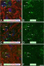
          Hibernating mammals undergo torpor periods characterized by a general decrease in body temperature, metabolic rate, and brain activity accompanied by complex adaptive brain changes that appear to protect the brain from extreme conditions of hypoxia and low temperatures. These processes are accompanied by morphological and neurochemical changes in the brain including those in cortical neurons such as the fragmentation and reduction of the Golgi apparatus (GA), which both reverse a few hours after arousal from the torpor state. In the present study, we characterized – by immunofluorescence and confocal microscopy – the GA of cortical astrocytes, oligodendrocytes, and microglial cells in the Syrian hamster, which is a facultative hibernator. We also show that after artificial induction of hibernation, in addition to neurons, the GA of glia in the Syrian hamster undergoes important structural changes, as well as modifications in the intensity of immunostaining and distribution patterns of Golgi structural proteins at different stages of the hibernation cycle.

          Related collections

          Most cited references54

          • Record: found
          • Abstract: found
          • Article: not found

          Characterization of a cis-Golgi matrix protein, GM130

          Antisera raised to a detergent- and salt-resistant matrix fraction from rat liver Golgi stacks were used to screen an expression library from rat liver cDNA. A full-length clone was obtained encoding a protein of 130 kD (termed GM130), the COOH-terminal domain of which was highly homologous to a Golgi human auto-antigen, golgin-95 (Fritzler et al., 1993). Biochemical data showed that GM130 is a peripheral cytoplasmic protein that is tightly bound to Golgi membranes and part of a larger oligomeric complex. Predictions from the protein sequence suggest that GM130 is an extended rod-like protein with coiled-coil domains. Immunofluorescence microscopy showed partial overlap with medial- and trans-Golgi markers but almost complete overlap with the cis-Golgi network (CGN) marker, syntaxin5. Immunoelectron microscopy confirmed this location showing that most of the GM130 was located in the CGN and in one or two cisternae on the cis-side of the Golgi stack. GM130 was not re-distributed to the ER in the presence of brefeldin A but maintained its overlap with syntaxin5 and a partial overlap with the ER- Golgi intermediate compartment marker, p53. Together these results suggest that GM130 is part of a cis-Golgi matrix and has a role in maintaining cis-Golgi structure.
            Bookmark
            • Record: found
            • Abstract: found
            • Article: not found

            Exiting the Golgi complex.

            The composition and identity of cell organelles are dictated by the flux of lipids and proteins that they receive and lose through cytosolic exchange and membrane trafficking. The trans-Golgi network (TGN) is a major sorting centre for cell lipids and proteins at the crossroads of the endocytic and exocytic pathways; it has a complex dynamic structure composed of a network of tubular membranes that generate pleiomorphic carriers targeted to different destinations. Live-cell imaging combined with three-dimensional tomography has recently provided the temporal and topographical framework that allows the assembly of the numerous molecular machineries so far implicated in sorting and trafficking at the TGN.
              Bookmark
              • Record: found
              • Abstract: found
              • Article: not found

              Reversible paired helical filament-like phosphorylation of tau is an adaptive process associated with neuronal plasticity in hibernating animals.

              Neurofibrillary pathology [paired helical filaments (PHFs)] formed by the microtubule-associated protein tau in a hyperphosphorylated form is a major hallmark of Alzheimer's disease and related disorders. The process of tau phosphorylation, thought to be of critical importance for PHF formation, and its potential link to neurodegeneration, however, is not understood very well, mostly because of the lack of a physiological in vivo model of PHF-like tau phosphorylation. Here we describe the formation of highly phosphorylated tau, containing a number of PHF-like epitopes in torpor during hibernation. PHF-like phosphorylation of tau was not associated with fibril formation and was fully reversible after arousal. Distribution of PHF-like tau followed a consistent pattern, being most intense in the entorhinal cortex, hippocampus, and isocortical areas. Within the hippocampus, a particularly high labeling was seen in CA3 pyramidal cells. Somewhat lesser reactivity was present in CA1 neurons while dentate gyrus granule cells were not reactive. Formation of PHF-like tau in CA3 neurons was paralleled by the regression of synaptic contacts of the mossy fiber system terminating on CA3 apical dendrites. Mossy fiber afferentation was re-established during arousal, concomitantly with the decrease of PHF-like tau in CA3 neurons. These findings implicate an essential link between neuronal plasticity and PHF-like phosphorylation of tau. The repeated formation and degradation of PHF-like tau might, thus, represent a physiological mechanism not necessarily associated with pathological effects. Hibernation will, therefore, be a valuable model to study the regulation of PHF-like tau-phosphorylation and its cell biological sequelae under physiological in vivo conditions.
                Bookmark

                Author and article information

                Contributors
                Journal
                Front Neuroanat
                Front Neuroanat
                Front. Neuroanat.
                Frontiers in Neuroanatomy
                Frontiers Media S.A.
                1662-5129
                19 November 2019
                2019
                : 13
                : 92
                Affiliations
                [1] 1Laboratorio Cajal de Circuitos Corticales (CTB), Universidad Politécnica de Madrid , Madrid, Spain
                [2] 2Departamento de Química y Bioquímica, Facultad de Farmacia, CEU San Pablo University, CEU Universities , Madrid, Spain
                [3] 3Instituto Cajal , CSIC, Madrid, Spain
                [4] 4Centro de Investigación Biomédica en Red de Enfermedades Neurodegenerativas , Madrid, Spain
                [5] 5Departamento de Biología Celular, Universidad Complutense de Madrid , Madrid, Spain
                Author notes

                Edited by: Thomas Arendt, Leipzig University, Germany

                Reviewed by: Joachim H. R. Lübke, Julich Research Centre, Germany; Fritz Geiser, University of New England, Australia

                *Correspondence: Gonzalo León-Espinosa, gonzalo.leonespinosa@ 123456ceu.es
                Article
                10.3389/fnana.2019.00092
                6882278
                c56c1f31-63e1-4131-adf2-3ed87cd0dc53
                Copyright © 2019 León-Espinosa, DeFelipe and Muñoz.

                This is an open-access article distributed under the terms of the Creative Commons Attribution License (CC BY). The use, distribution or reproduction in other forums is permitted, provided the original author(s) and the copyright owner(s) are credited and that the original publication in this journal is cited, in accordance with accepted academic practice. No use, distribution or reproduction is permitted which does not comply with these terms.

                History
                : 29 January 2019
                : 28 October 2019
                Page count
                Figures: 4, Tables: 1, Equations: 0, References: 60, Pages: 11, Words: 0
                Funding
                Funded by: Ministerio de Ciencia Tecnología y Telecomunicaciones 10.13039/501100006473
                Categories
                Neuroanatomy
                Original Research

                Neurosciences
                microglia,astrocytes,oligodendrocytes,torpor,gm130,golgin84,mg160
                Neurosciences
                microglia, astrocytes, oligodendrocytes, torpor, gm130, golgin84, mg160

                Comments

                Comment on this article