5
views
0
recommends
+1 Recommend
0 collections
    0
    shares
      • Record: found
      • Abstract: found
      • Article: found
      Is Open Access

      Effect of Phosphorylated Tau on Cortical Pyramidal Neuron Morphology during Hibernation

      research-article

      Read this article at

      Bookmark
          There is no author summary for this article yet. Authors can add summaries to their articles on ScienceOpen to make them more accessible to a non-specialist audience.

          Abstract

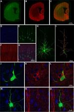
          The dendritic spines of pyramidal cells are the main postsynaptic target of excitatory glutamatergic synapses. Morphological alterations have been described in hippocampal dendritic spines during hibernation—a state of inactivity and metabolic depression that occurs via a transient neuronal tau hyperphosphorylation. Here, we have used the hibernating Syrian hamster to investigate the effect of hyperphosphorylated tau regarding neocortical neuronal structure. In particular, we examined layer Va pyramidal neurons. Our results indicate that hibernation does not promote significant changes in dendritic spine density. However, tau hyperphosphorylated neurons show a decrease in complexity, an increase in the tortuosity of the apical dendrites, and an increase in the diameter of the basal dendrites. Tau protein hyperphosphorylation and aggregation have been associated with loss or alterations of dendritic spines in neurodegenerative diseases, such as Alzheimer’s disease (AD). Our results may shed light on the correlation between tau hyperphosphorylation and the neuropathological processes in AD. Moreover, we observed changes in the length and area of the apical and basal dendritic spines during hibernation regardless of tau hyperphosphorylation. The morphological changes observed here also suggest region specificity, opening up debate about a possible relationship with the differential brain activity registered in these regions in previous studies.

          Related collections

          Most cited references69

          • Record: found
          • Abstract: found
          • Article: not found

          Correlation of Alzheimer disease neuropathologic changes with cognitive status: a review of the literature.

          Clinicopathologic correlation studies are critically important for the field of Alzheimer disease (AD) research. Studies on human subjects with autopsy confirmation entail numerous potential biases that affect both their general applicability and the validity of the correlations. Many sources of data variability can weaken the apparent correlation between cognitive status and AD neuropathologic changes. Indeed, most persons in advanced old age have significant non-AD brain lesions that may alter cognition independently of AD. Worldwide research efforts have evaluated thousands of human subjects to assess the causes of cognitive impairment in the elderly, and these studies have been interpreted in different ways. We review the literature focusing on the correlation of AD neuropathologic changes (i.e. β-amyloid plaques and neurofibrillary tangles) with cognitive impairment. We discuss the various patterns of brain changes that have been observed in elderly individuals to provide a perspective for understanding AD clinicopathologic correlation and conclude that evidence from many independent research centers strongly supports the existence of a specific disease, as defined by the presence of Aβ plaques and neurofibrillary tangles. Although Aβ plaques may play a key role in AD pathogenesis, the severity of cognitive impairment correlates best with the burden of neocortical neurofibrillary tangles.
            Bookmark
            • Record: found
            • Abstract: found
            • Article: not found

            Synapse loss and microglial activation precede tangles in a P301S tauopathy mouse model.

            Filamentous tau inclusions are hallmarks of Alzheimer's disease (AD) and related tauopathies, but earlier pathologies may herald disease onset. To investigate this, we studied wild-type and P301S mutant human tau transgenic (Tg) mice. Filamentous tau lesions developed in P301S Tg mice at 6 months of age, and progressively accumulated in association with striking neuron loss as well as hippocampal and entorhinal cortical atrophy by 9-12 months of age. Remarkably, hippocampal synapse loss and impaired synaptic function were detected in 3 month old P301S Tg mice before fibrillary tau tangles emerged. Prominent microglial activation also preceded tangle formation. Importantly, immunosuppression of young P301S Tg mice with FK506 attenuated tau pathology and increased lifespan, thereby linking neuroinflammation to early progression of tauopathies. Thus, hippocampal synaptic pathology and microgliosis may be the earliest manifestations of neurodegenerative tauopathies, and abrogation of tau-induced microglial activation could retard progression of these disorders.
              Bookmark
              • Record: found
              • Abstract: found
              • Article: not found

              Tau in physiology and pathology.

              Tau is a microtubule-associated protein that has a role in stabilizing neuronal microtubules and thus in promoting axonal outgrowth. Structurally, tau is a natively unfolded protein, is highly soluble and shows little tendency for aggregation. However, tau aggregation is characteristic of several neurodegenerative diseases known as tauopathies. The mechanisms underlying tau pathology and tau-mediated neurodegeneration are debated, but considerable progress has been made in the field of tau research in recent years, including the identification of new physiological roles for tau in the brain. Here, we review the expression, post-translational modifications and functions of tau in physiology and in pathophysiology.
                Bookmark

                Author and article information

                Contributors
                Journal
                Cereb Cortex Commun
                Cereb Cortex Commun
                cercorcomms
                Cerebral Cortex Communications
                Oxford University Press
                2632-7376
                2020
                21 May 2020
                21 May 2020
                : 1
                : 1
                : tgaa018
                Affiliations
                Laboratorio Cajal de Circuitos Corticales , Centro de Tecnología Biomédica , Universidad Politécnica de Madrid, Madrid 28002, Spain
                Laboratorio Cajal de Circuitos Corticales , Centro de Tecnología Biomédica , Universidad Politécnica de Madrid, Madrid 28002, Spain
                Instituto Cajal , Consejo Superior de Investigaciones Científicas, Madrid 28002, Spain
                Laboratorio Cajal de Circuitos Corticales , Centro de Tecnología Biomédica , Universidad Politécnica de Madrid, Madrid 28002, Spain
                Laboratorio Cajal de Circuitos Corticales , Centro de Tecnología Biomédica , Universidad Politécnica de Madrid, Madrid 28002, Spain
                Instituto Cajal , Consejo Superior de Investigaciones Científicas, Madrid 28002, Spain
                Laboratorio Cajal de Circuitos Corticales , Centro de Tecnología Biomédica , Universidad Politécnica de Madrid, Madrid 28002, Spain
                Facultad de Farmacia , Departamento de Química y Bioquímica, Universidad San Pablo-CEU , CEU Universities, Urbanización Montepríncipe, Boadilla del Monte, Madrid 28660, Spain
                Author notes
                Address correspondence to Gonzalo León-Espinosa, Facultad de Farmacia, Departamento de Química y Bioquímica, Universidad San Pablo-CEU, CEU Universities, Urbanización Montepríncipe, Boadilla del Monte, Madrid 28660, Spain. Email: gonzalo.leonespinosa@ 123456ceu.es .
                Author information
                http://orcid.org/0000-0002-9921-2614
                Article
                tgaa018
                10.1093/texcom/tgaa018
                8152943
                34296096
                7182692c-81e5-4748-ac43-fd8188ab9321
                © The Author(s) 2020. Published by Oxford University Press.

                This is an Open Access article distributed under the terms of the Creative Commons Attribution License ( http://creativecommons.org/licenses/by/4.0/), which permits unrestricted reuse, distribution, and reproduction in any medium, provided the original work is properly cited.

                History
                : 2 May 2020
                : 2 May 2020
                : 3 May 2020
                Page count
                Pages: 25
                Funding
                Funded by: Ministerio de Ciencia, Innovación y Universidades, DOI 10.13039/100014440;
                Award ID: PGC2018-094307-B-I00
                Funded by: Cajal Blue Brain Project;
                Funded by: Centro de Investigación en Red sobre Enfermedades Neurodegenerativas;
                Award ID: CB06/05/0066
                Categories
                Original Article

                3d reconstructions,cerebral cortex,dendrites,dendritic spines,syrian hamster

                Comments

                Comment on this article